


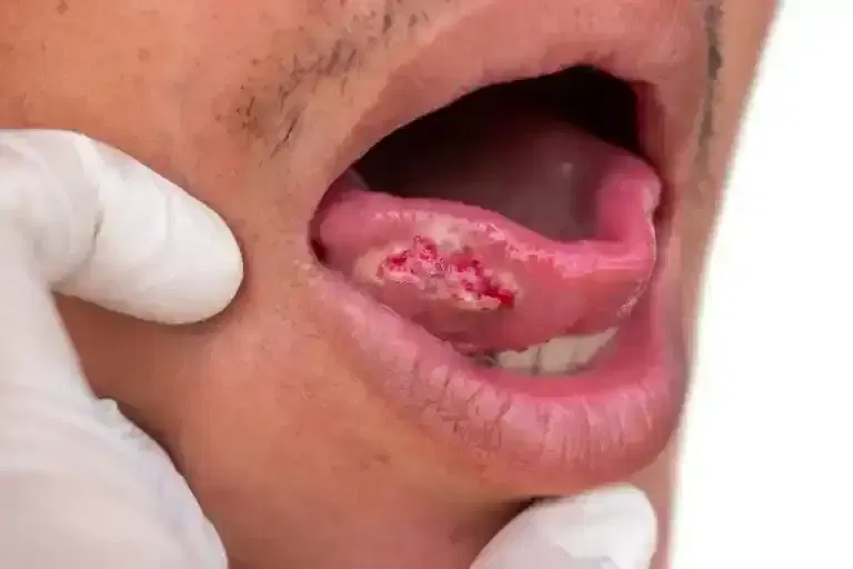
oral cancer surgery
5K+ International Patients Treated
40+ Source Countries Served
500+ Accredited Partner Hospitals
98% Patient Satisfaction
80% Average Savings vs USA
10K+ Doctors
NABH, JCI Accredited Hospitals
Free Treatment Plan
Free Consultation with Doctor
5+ Destinations Covered
About oral cancer surgery
Sources and Guidelines Referenced
This clinical guide synthesizes recommendations, consensus statements, and evidence from leading head and neck oncology societies and peer-reviewed studies:
- NCCN Guidelines: National Comprehensive Cancer Network Clinical Practice Guidelines in Oncology: Head and Neck Cancers (Version 2.2024).
- ASCO Guidelines: American Society of Clinical Oncology Practice Guideline on Management of the Neck in Squamous Cell Carcinoma of the Oral Cavity (2021).
- AHNS Guidelines: American Head and Neck Society Consensus Statement on Surgical Margins in Head and Neck Cancer (2022).
- ESMO Guidelines: European Society for Medical Oncology Clinical Practice Guidelines for Squamous Cell Carcinoma of the Head and Neck (2020).
- NICE Guidelines: National Institute for Health and Care Excellence Guideline NG36: Cancer of the upper aerodigestive tract (2016, updated 2021).
- Amit et al. (2013): Journal of Clinical Oncology, study on clear margin thresholds and survival in oral cavity cancer.
Oral Cancer Surgery: A Comprehensive Patient Guide
1. Definition and Medical Identity
Oral cancer surgery refers to the operative resection of malignant tumors originating within the oral cavity, including the tongue, lips, gums, floor of the mouth, and hard palate. Performed by head and neck surgical oncologists, its primary clinical goal is achieving complete tumor elimination with clear surgical margins while preserving critical speech, swallowing, and airway functions.
Surgical intervention remains the primary treatment approach for the majority of oral cavity cancers. The technical name for the removal of the primary tumor varies based on anatomical site and extent: partial glossectomy (removal of part of the tongue), glossectomy (complete tongue removal), mandibulectomy (removal of part or all of the lower jawbone), or maxillectomy (removal of the upper jawbone). When combined with regional lymph node removal, the procedure includes a neck dissection. Tissue repair performed at the same time is termed immediate reconstructive surgery, frequently utilizing microvascular free flap transfer.
2. The Underlying Condition or Need
Oral cancer surgery treats malignant cellular transformations within the mucosal lining of the oral cavity. Over 90% of oral malignancies are oral squamous cell carcinoma (OSCC), arising from the flat squamous cells that cover the mouth surface. Left untreated, these lesions progressively invade deeper muscle, nerve, and bone tissues, impairing essential functions and spreading to cervical lymph nodes.
The disease typically presents as a non-healing ulcer, a red patch (erythroplakia), a white patch (leukoplakia), or a painful mass within the mouth. Chronic irritation from tobacco smoke, heavy alcohol consumption, betel nut chewing, and human papillomavirus (HPV) infection are established oncogenic drivers (NCCN 2024). If untreated, oral carcinoma follows an aggressive local trajectory, eroding the jawbone, causing severe pain, preventing oral nutrition, compromising the upper airway, and metastasizing to regional neck nodes and distant organs such as the lungs and liver.
3. How the Treatment Works — Mechanism
Oral cancer surgery works by physically removing the entire malignant cell population from the patient's body along with a safety buffer of healthy surrounding tissue. By excising the primary tumor and involved regional lymph channels, surgery eliminates the source of disease before it can metastasize further or cause catastrophic local destruction.
At the cellular level, cancer cells exhibit invasive growth into surrounding tissue planes, blood vessels, and nerve sheath spaces (termed perineural invasion). Surgeons utilize physical excision guided by intraoperative frozen section biopsy—a microscopic laboratory analysis performed during the operation—to verify that no microscopic malignant cells remain at the cut edges (surgical margins). Removing regional lymph nodes via neck dissection stops the progression of regional lymphatic spread. Immediate microvascular reconstruction restores structural integrity by transferring well-vascularized tissue (skin, muscle, and bone) from another body site, reconnecting tiny blood vessels under a microscope to ensure flap survival.
4. Types and Variations
Surgeons categorize oral cancer procedures based on the tumor location, depth of invasion, involvement of bony structures, and regional lymphatic risk. Early-stage tumors may require localized soft-tissue excision, whereas advanced tumors demand composite resections involving soft tissue, bone, lymph nodes, and complex microvascular reconstruction.
The selection of surgical approach depends on clinical staging according to the AJCC Cancer Staging Manual (8th Edition), which incorporates tumor size, depth of invasion (DOI), and regional node status. The table below details the primary surgical procedures utilized in oral cavity cancer management.
| Surgical Procedure | Target Structures Removed | Primary Clinical Indication | Reconstructive Requirement |
|---|---|---|---|
| Wide Local Excision | Primary tumor + 1 cm soft tissue margin | Early-stage (T1-T2) superficial mucosal lesions | Primary closure or local mucosal flap |
| Partial / Subtotal Glossectomy | Portion of mobile tongue soft tissue | Squamous cell carcinoma of the lateral tongue | Primary closure, skin graft, or soft tissue flap |
| Marginal Mandibulectomy | Rim of jawbone, preserving bony continuity | Tumors adjacent to or minimally eroding mandibular periosteum | Gingival tissue flap or soft tissue coverage |
| Segmental Mandibulectomy | Full-thickness segment of lower jawbone | Tumors invading deep cortical jawbone structure | Vascularized bone free flap (e.g., fibula) |
| Selective Neck Dissection | Lymph node levels I–III (or I–IV) | Clinically node-negative neck (N0) with DOI > 3mm | None (cervical skin incision closure) |
| Comprehensive Neck Dissection | Lymph node levels I–V ± non-lymphatic structures | Clinically positive neck nodes (N+) or extensive nodal disease | None or muscular transposition flap |
5. Who the Treatment Is For — Indications
Oral cancer surgery is indicated as the primary definitive therapy for resectable malignant tumors of the oral cavity. According to NCCN Guidelines (2024), primary surgery is the treatment of choice for Stage I through Stage IVA oral squamous cell carcinoma due to superior oncologic control and lower functional morbidity compared to primary radiation.
Key clinical indications and patient eligibility criteria include:
- Histologically Proven Malignancy: Biopsy-confirmed squamous cell carcinoma, adenocarcinoma, or minor salivary gland carcinoma of the oral cavity.
- Resectable Tumors: Disease that can be fully excised with clear margins without causing unacceptable functional impairment or mortality.
- Depth of Invasion Threshold: Tumors with a depth of invasion greater than 3 millimeters warrant elective neck dissection, even in the absence of clinically detectable lymph node swelling (ASCO 2021).
- Bone Involvement: Radiographic or clinical evidence of jawbone erosion requiring marginal or segmental bone resection.
- Pre-Malignant Lesions: High-grade severe dysplasia or carcinoma in situ failing conservative non-surgical management.
6. Who the Treatment Is NOT For — Contraindications
Oral cancer surgery is not suitable for patients with unresectable local disease, widespread distant metastases, or severe medical comorbidities that make general anesthesia life-threatening. In these scenarios, non-surgical modalities such as systemic therapy or palliative radiation are preferred.
Specific absolute and relative contraindications include:
- Unresectable Disease (Absolute): Fixation of the tumor to the carotid artery, prevertebral fascia, or skull base, where complete resection with clear margins is physically impossible (NCCN 2024).
- Distant Metastasis (Relative/Absolute): Presence of widespread metastatic disease (Stage IVC) in lungs, liver, or bone, where major curative resection does not offer a survival benefit. Palliative local surgery may occasionally be considered.
- Severe Cardiopulmonary Dysfunction: Severe heart failure, recent myocardial infarction, or severe end-stage lung disease precluding lengthy general anesthesia.
- Uncontrolled Systemic Coagulopathy: Inability to safely undergo major surgery due to uncorrectable bleeding disorders.
7. Alternatives and Clinical Comparison
The main alternative to primary oral cancer surgery is definitive radiotherapy, often combined with concurrent chemotherapy or targeted therapy. However, clinical evidence strongly favors surgery for oral cavity tumors, as bone and mucosal tissues in the mouth are highly sensitive to high-dose radiation damage (ESMO 2020).
Unlike oropharyngeal cancers (such as tonsil or base of tongue tumors), which are frequently driven by HPV and respond favorably to primary chemoradiation, oral cavity cancers are predominantly smoking/alcohol-related and demonstrate lower radiocurability. The table below outlines how primary surgery compares with alternative treatment strategies.
| Treatment Modality | Primary Mechanism | Key Advantages | Primary Limitations & Trade-offs |
|---|---|---|---|
| Primary Surgery | Physical excision of tumor and affected lymph nodes | High local control; exact pathologic margin assessment; avoids jaw osteoradionecrosis | Requires surgical recovery; potential altered speech, swallowing, and cosmetic changes |
| Definitive Radiotherapy | High-energy ionizing radiation damaging cancer cell DNA | Preserves anatomical tissue structures without surgical incisions | Risk of osteoradionecrosis of jawbone; chronic dry mouth; lower local cure rates for OSCC |
| Concurrent Chemoradiation | Radiation combined with radiosensitizing chemotherapy (e.g., Cisplatin) | Combines local and systemic attack for locally advanced disease | Significant acute mucosal toxicity; severe long-term swallowing difficulty; systemic side effects |
| Systemic Immunotherapy / Chemo | PD-1 inhibitors (Pembrolizumab) or systemic cytotoxic drugs | Treats widespread systemic or recurrent non-resectable disease | Palliative intent; rarely yields complete durable cure as monotherapy in advanced OSCC |
8. Pre-Treatment Phase
The pre-treatment phase establishes diagnostic precision, staging, functional baseline, and multidisciplinary operative planning. Patients undergo comprehensive imaging, dental evaluation, and systemic medical optimization over 1 to 3 weeks prior to surgery.
Diagnostic workup begins with a detailed physical examination, direct endoscopic evaluation, and a tissue biopsy. High-resolution contrast-enhanced CT or MRI scans of the head and neck are performed to evaluate tumor extent, soft tissue invasion, depth of invasion, and lymph node status. A panorex dental X-ray or dedicated jaw CT assesses bone invasion. PET-CT imaging is recommended for Stage III–IV disease to rule out distant metastases (NCCN 2024).
Mandatory pre-operative preparation steps include:
- Multidisciplinary Tumor Board Review: Case review involving surgical oncologists, radiation oncologists, medical oncologists, radiologists, pathologists, and speech pathologists.
- Comprehensive Dental Clearance: Extraction of unresolvably decayed or infected teeth within planned surgical or future radiation fields to prevent post-treatment osteoradionecrosis.
- Functional Baseline Testing: Evaluation by a speech and language pathologist to record baseline speech clarity and swallowing function.
- Medical Optimization: Cardiopulmonary risk stratification, cessation of blood-thinning medications, and complete cessation of alcohol and tobacco use.
9. The Procedure — Step-by-Step Clinical Detail
Oral cancer surgery is a major inpatient procedure conducted under general anesthesia in a main operating theater. Depending on the complexity of resection and microvascular reconstruction, the surgical duration ranges from 3 to 12 hours.
The step-by-step clinical progression involves six main operational phases:
- Phase 1: Anesthesia and Airway Securing: General anesthesia is administered. The surgical team secures the airway via nasal intubation or by performing a temporary tracheostomy to ensure safe ventilation throughout surgery and early recovery.
- Phase 2: Tumor Resection: The head and neck surgeon excises the primary oral tumor with a planned 1.0 to 1.5 cm visible margin to achieve a final microscopic clear margin of at least 5 mm. Bone resection (marginal or segmental mandibulectomy) is executed if bone involvement is present.
- Phase 3: Frozen Section Analysis: Tissue samples from the resection margins are immediately sent to the pathology laboratory for frozen section analysis. If tumor cells are identified at any edge, additional tissue is removed until all margins are declared negative.
- Phase 4: Neck Dissection: A cervical incision is made to access the neck. The surgeon systematically removes lymph-node-bearing fatty tissue from designated cervical levels (I through V) while preserving vital nerves (such as the vagus and spinal accessory nerves) and major blood vessels whenever safely possible.
- Phase 5: Microvascular Free Flap Reconstruction: If a significant soft tissue or bone defect remains, a reconstructive team harvests tissue from another body site (such as skin and muscle from the radial forearm or bone from the fibula). Using an operating microscope, the flap's artery and vein are anastomosed (reconnected) to target vessels in the neck to restore tissue blood flow.
- Phase 6: Closure and Drain Placement: Suction drains are positioned in the neck to prevent fluid accumulation (seroma/hematoma). Surgical incisions are closed in layers using sutures and skin staples. Feeding tubes (nasogastric or gastrostomy) are confirmed in position.
10. Immediate Post-Procedure Period
In the first 24 to 48 hours following oral cancer surgery, intensive clinical monitoring focuses on airway maintenance, microvascular flap viability checks, fluid management, and acute pain control. Patients are routinely cared for in an Intensive Care Unit (ICU) or specialized Head and Neck Surgical Step-Down Unit.
Essential immediate post-operative protocols include:
- Airway Management: Tracheostomy tube care involves frequent suctioning, humidification, and inner cannula cleaning to maintain airway patency.
- Flap Viability Monitoring: If free flap reconstruction was performed, nurses conduct hourly evaluations of flap color, capillary refill, temperature, and Doppler ultrasound arterial/venous signals to detect blood vessel thrombosis early.
- Pain Control: Intravenous patient-controlled analgesia (PCA) or multimodal pain protocols combining opioids, acetaminophen, and neuropathic medications maintain comfort without suppressing respiratory drive.
- Enteral Nutrition: Oral intake is restricted to allow oral mucosal incisions to heal without contamination. Nutrition and medications are delivered through a nasogastric (NG) tube or percutaneous endoscopic gastrostomy (PEG) tube.
11. Recovery — Short and Long Term
Recovery from oral cancer surgery is a staged process involving acute surgical wound healing, inpatient mobilization, outpatient functional rehabilitation, and long-term oncologic surveillance. Total hospital length of stay averages 5 to 10 days.
The post-operative timeline unfolds across distinct recovery phases:
- Inpatient Days 3–7: Patients transition from intravenous to enteral or oral medications. Drains are removed when output drops below 30 mL/day. Physical therapy assists with early ambulation. Speech and swallowing therapists initiate gentle therapy protocols.
- Weeks 2–4 (Discharge & Initial Outpatient Recovery): Suture and staple removal occurs. Tracheostomy tubes are typically decannulated (removed) once airway swelling subsides. Patients receiving free flaps continue wound care. Adjuvant therapy recommendations are finalized based on pathology reports.
- Months 1–3 (Adjuvant Treatment & Rehabilitation): If high-risk pathology features are present, adjuvant radiation or chemoradiation starts within 6 weeks of surgery. Intensive swallowing exercises, mouth-opening exercises (to prevent jaw stiffness/trismus), and speech therapy continue weekly.
- Months 3–12 (Long-Term Adaptation): Swallowing and speech capabilities stabilize. Dental rehabilitation (such as dental implants or specialized dentures) may begin once tissue healing and radiation protocols are complete. Serial clinical examinations and surveillance neck imaging occur every 2 to 3 months.
12. Risks, Side Effects, and Complications
Oral cancer surgery carries risks inherent to major head and neck surgical procedures. Complications vary based on resection extent, bone involvement, patient medical status, and microvascular reconstructive complexity.
Surgeons stratify potential adverse events into mild/common side effects, moderate complications, and rare/severe adverse outcomes. The table below provides a comprehensive risk severity matrix.
| Severity Level | Complication / Adverse Event | Clinical Presentation & Management |
|---|---|---|
| Common / Mild | Local edema, temporary dysphagia, mild facial pain, temporary numbness of lip/tongue | Expected post-operative occurrences; managed with analgesics, elevation, and therapy. |
| Uncommon / Moderate | Surgical site infection, wound dehiscence, hematoma, salivary fistula, shoulder dysfunction | Requires antibiotic therapy, localized wound care, drainage, or physical therapy for nerve irritation. |
| Rare / Severe | Free flap thrombosis/failure, carotid artery rupture, deep vein thrombosis (DVT), pulmonary embolism | Medical emergencies requiring immediate surgical re-exploration, anticoagulation, or critical resuscitation. |
Severe long-term complications can include trismus (severe restriction in jaw opening due to scar tissue or radiation) and permanent altered sensation in the lower lip and tongue due to surgical sacrifice or manipulation of the inferior alveolar or lingual nerves. If post-operative radiation is administered, long-term xerostomia (chronic dry mouth) and increased risk of dental caries may occur.
13. Lifestyle and Behavioural Considerations
Modifying lifestyle habits before and after oral cancer surgery directly impacts surgical healing, free flap survival, functional recovery, and long-term overall survival. Absolute cessation of tobacco and alcohol is the most crucial lifestyle factor.
Key behavioural considerations include:
- Tobacco and Alcohol Cessation: Continued smoking impairs surgical wound healing, doubles the risk of microvascular flap failure, and increases the rate of secondary primary head and neck cancers by up to 30% (ASCO 2021). Smoking cessation interventions must begin immediately upon diagnosis.
- Nutritional Optimization: Malnutrition impairs tissue healing and increases infection risk. High-calorie, high-protein liquid supplements delivered via feeding tubes maintain body mass during recovery until safe oral swallowing resumes.
- Oral Hygiene Maintenance: Post-operative oral rinses (such as dilute salt and baking soda solutions or chlorhexidine) keep mucosal incisions clean and reduce bacterial colonization without irritating delicate healing tissue.
- Jaw Stretching Exercises: Daily mouth-opening exercises using specialized jaw-stretching devices or stacked tongue depressors prevent permanent jaw stiffness (trismus) caused by surgical scarring.
14. How Outcomes Are Measured
Clinical success in oral cancer surgery is evaluated using three primary endpoints: oncologic disease control, overall survival, and functional restoration of speech and swallowing. Standardized pathologic criteria determine whether surgical goals have been met.
Oncologic control is measured primarily by margin status and regional disease control:
- Surgical Margin Status: A clear margin (> 5 mm of healthy tissue microscopically) represents successful excision. Close margins (1–5 mm) or positive margins (< 1 mm or tumor at the cut edge) indicate a higher risk of local recurrence and require adjuvant radiation or re-excision (AHNS 2022).
- Pathologic Risk Stratification: Features such as extranodal extension (ENE)—where cancer breaks through the lymph node capsule—and positive margins are major high-risk indicators requiring post-operative chemoradiation (NCCN 2024).
- Overall and Disease-Free Survival: Early-stage (Stage I–II) oral cavity cancers treated with primary surgery achieve 5-year overall survival rates between 70% and 90%, whereas locally advanced stages (Stage III–IV) yield 5-year survival rates ranging from 30% to 60%, depending on nodal status and ENE (Amit et al., 2013).
- Functional Endpoints: Standardized scales evaluate swallowing safety (prevention of aspiration), speech intelligibility, and quality of life at 3, 6, and 12 months post-surgery.
15. Recent Advances and Current Standard of Care
Over the past 15 years, the standard of care for oral cancer surgery has advanced significantly through innovations in computer-assisted surgical planning, minimally invasive techniques, microvascular reconstructive techniques, and targeted adjuvant therapies.
Key modern clinical advances include:
- Virtual Surgical Planning (VSP) & 3D Printing: Pre-operative CT scans are used to design custom 3D cutting guides and pre-bent titanium plates. This technology allows surgeons to perform highly precise jaw bone resections and fibula free flap shaping, reducing operative time and improving cosmetic and functional alignment.
- Sentinel Lymph Node Biopsy (SLNB): For select early-stage (T1–T2 N0) oral cancers, SLNB utilizes radiotracer mapping to identify and remove only the primary draining lymph nodes, avoiding a full neck dissection if nodes are clear (ASCO 2021).
- Advanced Free Flap Reconstruction: Perforator-based free flaps (such as the anterolateral thigh flap) allow surgeons to harvest tissue with minimal donor-site muscle damage, improving donor-site recovery.
- Integration of Immunotherapy: Clinical trials are evaluating neoadjuvant (pre-operative) immunotherapy with PD-1 checkpoint inhibitors (e.g., pembrolizumab) to shrink advanced tumors prior to surgery and improve systemic immune responses (NCCN 2024).
16. Common Myths and Misconceptions
Misinformation regarding oral cancer surgery often causes unnecessary anxiety and delays in seeking definitive medical care. The following evidence-based corrections address common misconceptions.
Myth: Surgery causes oral cancer to spread faster by exposing tumor cells to air.
Reality: Surgical excision does not cause cancer to spread. Surgery is performed using precise oncologic techniques to completely remove malignant tissue with clear margins, directly lowering recurrence rates (NCCN 2024).
Myth: Having oral cancer surgery means losing the ability to speak permanently.
Reality: While tongue or jaw surgery alters speech articulation initially, modern microvascular reconstruction and dedicated speech rehabilitation allow the majority of patients to regain understandable, functional speech.
Myth: Radiation therapy is always better than surgery because it avoids facial incisions.
Reality: For oral cavity cancers, primary surgery delivers higher local cure rates and avoids major complications like bone radiation necrosis (osteoradionecrosis) when compared to primary radiation (ESMO 2020).
Myth: Neck dissection will result in permanent complete paralysis of the neck and arm.
Reality: Modern selective neck dissections preserve non-lymphatic structures such as the spinal accessory nerve whenever safely possible, minimizing shoulder weakness and neck stiffness.
Myth: If lymph nodes in the neck are removed, the body can no longer drain lymphatic fluid from the head.
Reality: The neck contains hundreds of lymph nodes arranged in parallel networks. Removing specific regional lymph node levels leaves remaining lymphatic pathways to handle drainage, though temporary mild lymphedema can occur.
Myth: Oral cancer only affects older people who smoke heavily.
Reality: While tobacco and alcohol are major risk factors, oral cancer is increasingly diagnosed in younger non-smoking individuals due to emerging genetic and environmental factors (ACS 2023).
17. Frequently Asked Questions
What is oral cancer surgery?
Oral cancer surgery is the operative removal of cancerous tumors originating in the mouth, including the tongue, gums, cheek lining, and jawbone. Performed by head and neck surgical oncologists, the procedure aims to excise the malignancy completely with clear margins while reconstructing involved structures to preserve essential functions like speech and swallowing.
How long does oral cancer surgery take?
The duration varies depending on surgical complexity. A wide local excision for an early-stage superficial tumor may take 1 to 2 hours. Complex composite resections involving jawbone removal, neck dissection, and microvascular free flap reconstruction typically require 6 to 12 hours of specialized operative time.
Will I have visible facial scars after oral cancer surgery?
Incisions are carefully planned along natural skin creases in the neck and lower lip whenever possible to minimize cosmetic visibility. Many resections are performed entirely through the open mouth (transorally). When external incisions are required for jaw access or neck dissection, scars fade significantly over 6 to 12 months.
How will I eat and drink after surgery?
Immediately after surgery, oral eating is paused to allow internal mouth incisions to heal safely. Nutrition is provided through a temporary nasogastric or gastrostomy feeding tube. Once the surgical team confirms safe healing and swallowing capability—usually within 1 to 2 weeks—patients gradually transition from liquid to soft and solid foods.
What is a neck dissection and why is it necessary?
A neck dissection is a surgical procedure that removes lymph nodes from specific levels of the neck. It is necessary because oral cavity cancers frequently spread to regional cervical lymph nodes. Removing these nodes treats microscopic metastatic disease and provides accurate pathological staging to guide further adjuvant treatments.
What is a free flap reconstruction?
Free flap reconstruction involves transferring healthy tissue—such as skin, muscle, or bone—from another part of the patient's body (like the forearm, thigh, or leg) to repair the surgical defect in the mouth. The flap's blood vessels are reattached to neck blood vessels under a microscope to ensure immediate, permanent tissue survival.
Will I need a temporary tracheostomy tube?
A temporary tracheostomy (a breathing tube inserted into the windpipe through a small neck incision) is often performed during major oral resections or jaw surgeries. It protects the airway from post-operative swelling in the mouth and throat. The tube is typically removed within 3 to 7 days as swelling resolves.
Is oral cancer surgery painful?
Patients experience moderate post-operative discomfort, which is effectively managed using intravenous analgesics, nerve blocks, and oral or tube-administered pain medications. Pain gradually decreases over the first 2 weeks. The medical team utilizes structured multimodal pain control protocols to keep patients comfortable.
How long will I stay in the hospital?
The typical hospital stay ranges from 5 to 10 days. Patients undergoing minor transoral resections may be discharged in 1 to 3 days, whereas patients requiring complex bone resections, free flap reconstruction, or tracheostomy care remain hospitalized longer for specialized monitoring and therapy.
When can I return to work and daily activities?
Most patients resume light daily activities within 3 to 4 weeks post-discharge. Returning to work depends on physical job demands, speech requirements, and whether adjuvant radiation or chemotherapy is needed. Patients receiving surgery alone often return to work in 4 to 6 weeks, while those undergoing radiation may require 3 to 6 months.
Will I need radiation or chemotherapy after surgery?
Adjuvant radiation or chemoradiation is recommended if final pathology reports reveal high-risk features, such as positive surgical margins, cancer spread into multiple lymph nodes, extranodal extension (ENE), or deep perineural invasion (NCCN 2024). Early-stage tumors excised with clear margins usually require surgery alone.
How does surgery affect speech and talking?
Speech changes depend on the location and amount of tissue removed, particularly from the tongue and palate. While initial speech may sound muffled due to swelling, reconstructive surgery and targeted speech therapy help patients adapt, allowing most to achieve clear, understandable speech within several months.
What are the signs of cancer recurrence after surgery?
Warning signs of local recurrence include a new non-healing mouth ulcer, a persistent red or white patch, new localized pain, a firm lump in the mouth or neck, difficulty swallowing, or unexplained weight loss. Patients undergo serial physical examinations and surveillance imaging every 2 to 3 months during early follow-up.
Booking With DIVINHEAL
Get a free consultation to understand your treatment options
Cost Calculator
I know my treatment - show me cost from 3 hospitals
Plan My Journey
Tell us your condition and budget - our AI matches the right destination, hospital and doctor and visa pathway
Recommended Article

Booking With DIVINHEAL
Get a free consultation to understand your treatment options
Cost Calculator
I know my treatment - show me cost from 3 hospitals
Plan My Journey
Tell us your condition and budget - our AI matches the right destination, hospital and doctor and visa pathway
Recommended Article
Our Speciality and Treatments
oculoplastic surgery
Macular Degeneration Treatment (AMD)
Pediatric Ophthalmology
Squint / Strabismus Surgery (Adult)
ICL / Phakic IOL Implantation
LASIK / SMILE / PRK (Refractive Surgery)
Keratoconus Treatment (CXL / Rings)
Corneal Transplant (Keratoplasty)
Pediatric Squint Surgery
Diabetic Retinopathy Laser / Anti-VEGF Injections
Vitrectomy
Retinal Detachment Repair
Pterygium Surgery
Glaucoma Surgery (Trabeculectomy / MIGS)
Cataract Surgery (Phaco with IOL)






Meet Our Medical Specialists
Hospitals
NABH & JCI Accredited Hospitals in India,Turkey, Thailand & UAE.
Hisar Intercontinental Hospital
Saray Mah. Siteyolu Cad. No:7, Umraniye, 34768, Istanbul, Turkey
Medical Park Group, Istanbul
Fahrettin Kerim Gokay Cad. Tıbbiye Cd., Kadikoy, Istanbul, Turkey
Emsey Hospital, Pendik, Istanbul
Çamlık, Selçuklu Cd. No:22, 34912 Pendik/İstanbul, Türkiye
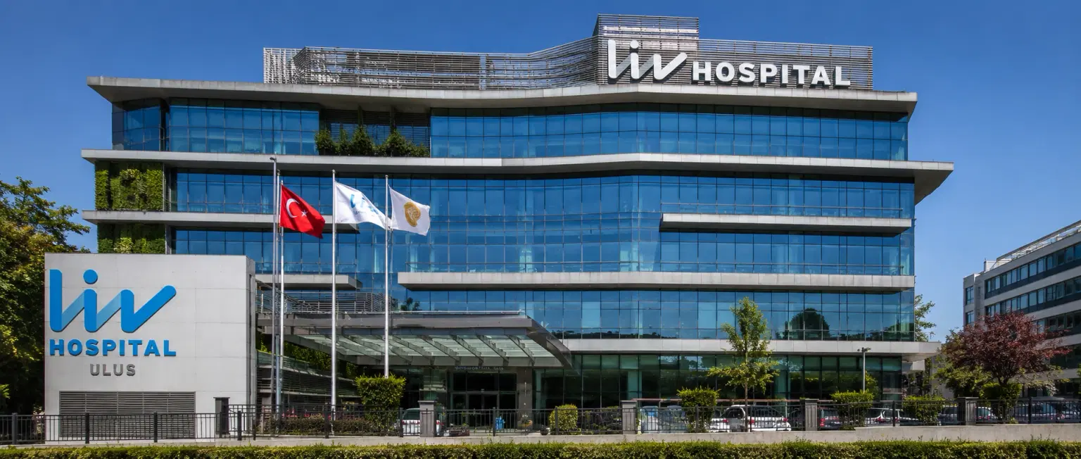
LIV Hospital, Istanbul
How DivinHeal Helps
We simplify your medical journey by providing comprehensive support and access to world-class healthcare.
Expert Specialist Matching
Connecting you with the world's top-rated medical experts.
Accredited Hospital Network
Access to JCI & NABH certified healthcare facilities.
Complete Travel Coordination
Hassle-free visa, stay, and local transport assistance.
24/7 Personal Care
Dedicated patient advisors supporting you at every step.
Journey Guidance
Full guidance from start to end of the patient treatment journey.
Expert Specialist Matching
Connecting you with the world's top-rated medical experts.
Everything you
need to know today
Browse through these common inquiries to better understand our patient-focused medical platform.
Yes, we work with a variety of insurance providers. Contact our team to verify your coverage.
Yes, we provide secure online consultations with experienced specialists.
Our care coordinators help match you with the most suitable specialist.
Absolutely. Your medical information is protected according to healthcare privacy standards.
Look at six things: accreditation (JCI or NABH), specialty depth, doctor credentials and experience, procedure-specific success rates, international patient support, and technology. DivinHeal's AI-driven matching evaluates every hospital in our accredited partner network on these dimensions and shortlists the best-fit options for your condition, budget, and country.
JCI (Joint Commission International) is the US-based global gold standard for hospital quality, recognised worldwide. NABH is India's national accreditation — accredited by ISQua, the same body that accredits JCI. Both signal independently verified safety and quality. Most of India's leading hospitals hold both.
Yes. All three welcome international patients through structured medical visa programs. India is the most established, treating patients from Africa, the Middle East, and South Asia at 60–80% lower cost. Thailand leads in cosmetic and dental care. The UAE is emerging in oncology and reproductive medicine.
Most patients save 50–80% on treatment costs. Heart bypass costs US $7,000–9,000 in India compared to $70,000–150,000 in the US. IVF costs $3,000–4,500 compared to $12,000–20,000 in the UK. Even after flights, visa, and accommodation, total savings remain 60–70%.
DivinHeal manages your entire non-medical journey: visa invitation letters, medical visa guidance, doctor appointments, teleconsultations, airport pickup, hospital-vetted accommodation for you and your attendant, language interpreters, local transport, cuisine preferences, and post-treatment follow-up — one dedicated coordinator from first enquiry to final follow-up.
You need a valid passport (6+ months validity), a medical visa (M-Visa for India — DivinHeal provides the hospital invitation letter), return flight tickets, recent medical reports and a doctor's referral, current prescription list, and proof of financial means. Any accompanying attendant needs their own passport and MX-Visa.
Still have more questions?
Book a call with our friendly team to learn how DivineHeal simplifies your healthcare journey.