



breast cancer surgery

5K+ International Patients Treated
40+ Source Countries Served
500+ Accredited Partner Hospitals
98% Patient Satisfaction
80% Average Savings vs USA
10K+ Doctors
NABH, JCI Accredited Hospitals
Free Treatment Plan
Free Consultation with Doctor
5+ Destinations Covered
About breast cancer surgery
Sources and Guidelines Referenced
National Comprehensive Cancer Network (NCCN) Clinical Practice Guidelines in Oncology: Breast Cancer (Version 2.2024); American Society of Clinical Oncology (ASCO) Endorsement of SSO/ASTRO Consensus Guidelines (2020); Society of Surgical Oncology (SSO) Guidelines on Margins for Breast-Conserving Surgery (2014, 2016); American College of Surgeons Oncology Group (ACOSOG) Z0011 Trial (Giuliano et al., 2017); St. Gallen International Consensus Conference Guidelines (2023); European Society for Medical Oncology (ESMO) Early Breast Cancer Clinical Practice Guidelines (2024).
Breast Cancer Surgery: A Comprehensive Patient Guide
1. Definition and Medical Identity
Breast cancer surgery is the surgical removal of cancerous tumor tissue from the breast along with associated tissue and regional lymph nodes. Known clinically as partial mastectomy, total mastectomy, or axillary surgery, it is a primary treatment modality in surgical oncology designed to remove primary tumors, prevent local disease spread, and guide staging.
Surgery remains the anchor of local control in multidisciplinary breast cancer care. The procedure entails removing the primary neoplasm—an abnormal growth of tissue—along with an adequate buffer of normal tissue called a margin. Depending on stage, biological subtype, and patient characteristics, surgery may precede or follow systemic treatments such as chemotherapy or hormone therapy.
2. The Underlying Condition or Need
Breast cancer surgery treats malignant transformations occurring within the epithelial cells of the breast lobules or ducts. Unchecked cellular proliferation leads to primary tumor formation, local tissue invasion, and potential spread to regional lymph nodes and distant organs. Surgery disrupts this progression by physically excising primary disease.
Most breast cancers present as non-palpable abnormalities detected on routine screening mammograms or as palpable breast masses. Without surgical intervention, primary malignant tumors typically continue to grow locally, causing tissue breakdown, skin involvement, severe localized pain, and increased risk of systemic distant metastasis. Surgical excision physically removes the burden of disease, allowing definitive tissue examination to guide long-term survival strategies.
3. How the Treatment Works — Mechanism
Breast cancer surgery achieves its goal by macroscopic physical excision of tumor tissue paired with pathological margin verification. The surgeon removes the malignant mass along with surrounding healthy tissue to ensure no microscopic cancer cells remain at the edge of the removed specimen.
At the anatomical level, surgery disrupts the local vascular and lymphatic pathways that support tumor growth. Lymph node evaluation—specifically identifying the sentinel lymph node (the primary receiving node for lymphatic drainage from the tumor area)—determines whether cancer cells have migrated into the lymphatic system. If the sentinel node is clear of cancer, further nodal surgery is avoided, preserving normal lymphatic drainage of the arm. Pathological analysis of excised tissue measures tumor size, histological grade, hormone receptor expression, and margin distance to inform subsequent adjuvant therapies.
4. Types and Variations
Breast cancer surgery is categorized into breast-conserving surgery and total tissue removal, combined with targeted or comprehensive axillary lymph node management. The choice depends on tumor size, location, breast size, tumor biology, and patient preferences according to NCCN guidelines.
Lumpectomy, also termed partial mastectomy or wide local excision, removes only the tumor mass and a small margin of surrounding normal breast tissue, leaving the majority of the breast intact. Mastectomy involves complete removal of the breast tissue, including the nipple-areola complex in standard total mastectomies, though skin-sparing and nipple-sparing techniques preserve outer skin envelopes for immediate reconstruction. Axillary evaluation involves either sentinel lymph node biopsy (SLNB) or formal axillary lymph node dissection (ALND).
| Surgical Type | Tissue Removed | Anatomical Preservation | Typical Invasiveness | Standard Indications |
|---|---|---|---|---|
| Lumpectomy (Breast-Conserving Surgery) | Tumor plus rim of normal surrounding tissue | Preserves majority of natural breast tissue | Low to Moderate | Unifocal early-stage tumors, favorable tumor-to-breast ratio |
| Total (Simple) Mastectomy | Entire breast tissue, fascia, skin, and nipple-areola complex | Removes entire breast parenchymal structure | Moderate | Multifocal disease, large tumor ratio, patient choice |
| Skin/Nipple-Sparing Mastectomy | Breast glandular tissue; preserves skin flap +/- nipple | Preserves outer skin envelope for reconstruction | Moderate to High | Early stage disease planning immediate breast reconstruction |
| Sentinel Lymph Node Biopsy (SLNB) | 1 to 4 primary drainage lymph nodes | Preserves deep axillary lymphatic channels | Low | Clinically node-negative invasive breast cancer |
| Axillary Lymph Node Dissection (ALND) | Level I and II axillary lymph node contents | Removes majority of axillary fat and nodes | High | Heavy nodal involvement, positive non-sentinel nodes |
5. Who the Treatment Is For — Indications
Breast cancer surgery is indicated for individuals diagnosed with non-invasive or invasive primary malignancies of the breast. Diagnostic confirmation requires pre-operative core needle biopsy, non-invasive imaging staging, and clinical evaluation to verify localized or regional disease amenable to resection.
Specific indications include:
- Ductal Carcinoma in Situ (DCIS): Non-invasive stage 0 disease requiring local excision to prevent progression to invasive carcinoma.
- Invasive Ductal or Lobular Carcinoma (Stages I–III): Primary invasive malignancies requiring definitive surgical clearing of the breast mass and axillary staging.
- Phyllodes Tumors: Rare fibroepithelial tumors requiring wide surgical margins to lower recurrence risks.
- Prophylactic (Risk-Reducing) Surgery: High-risk genetic mutation carriers (e.g., BRCA1, BRCA2, PALB2) selecting bilateral total mastectomy to minimize primary cancer risk.
- Local Recurrence: Recurrent localized disease following prior breast-conserving treatment.
6. Who the Treatment Is NOT For — Contraindications
Breast cancer surgery has specific absolute and relative contraindications based on systemic cancer spread, anatomical factors, or patient underlying health. Surgical decisions must balance local oncologic control against overall systemic health and operative safety.
Contraindications and limiting factors include:
- De Novo Stage IV Metastatic Disease: Primary surgery on the breast tumor is generally not indicated as initial therapy when distant organ metastases are present, except for systemic control or palliative symptom management (NCCN 2024).
- Inability to Achieve Negative Margins with Breast Conservation: Diffuse malignant-appearing microcalcifications across multiple quadrants or persistent positive surgical margins contraindicate continued lumpectomy, necessitating total mastectomy.
- Contraindications to Post-Operative Radiation: Prior chest wall radiation or active systemic connective tissue disorders (e.g., scleroderma) prohibit lumpectomy, as adjuvant radiation is mandatory after breast conservation.
- Severe Uncontrolled Medical Comorbidities: Advanced cardiac failure, severe chronic obstructive pulmonary disease, or acute systemic infection rendering general anesthesia unacceptably unsafe.
7. Alternatives and Clinical Comparison
Definitive management of localized breast cancer typically requires surgical removal. However, primary non-surgical modalities—such as systemic therapy alone—or different combinations of local modalities exist for specific clinical scenarios under established oncologic protocols.
In patients with hormone receptor-positive early breast cancer who are unfit for general anesthesia due to severe medical comorbidities, primary endocrine therapy (such as aromatase inhibitors) may control localized disease. Primary radiation therapy without surgery is rarely standard practice except in non-operable palliative settings. The table below compares primary local and systemic management strategies.
| Treatment Strategy | Mechanism of Action | Primary Clinical Context | Pros | Cons / Trade-offs |
|---|---|---|---|---|
| Breast Cancer Surgery (Standard) | Physical excision of primary tumor and staging nodes | Curative intent for Stages 0–III breast cancer | Definitive tissue clearing, precise staging, proven survival benefit | Surgical risks, anatomical alterational requirements |
| Neoadjuvant Systemic Therapy (Pre-op) | Pre-operative systemic tumor downsizing via chemotherapy/HER2 drugs | Large tumors, HER2+, or triple-negative disease prior to planned surgery | Downstages tumor, allows lumpectomy, tests drug response in vivo | Delays immediate surgery, risks non-response (rare) |
| Primary Endocrine Therapy (No Surgery) | Hormonal suppression of tumor cell proliferation | Frail, elderly, or severely medically unfit patients with ER+ tumors | Avoids surgical and anesthetic risks completely | Lower rates of complete local control; non-curative in long term |
8. Pre-Treatment Phase
The pre-treatment phase optimizes patient health, establishes clear anatomical boundaries of the malignancy, and formulates an individualized surgical plan. Multi-disciplinary input from surgical oncologists, medical oncologists, radiologists, and pathologists ensures alignment with current consensus guidelines.
Diagnostic evaluation includes diagnostic mammography, targeted ultrasound, and occasionally breast magnetic resonance imaging (MRI) to assess disease extent. Tissue confirmation via image-guided core needle biopsy establishes histological type and biomarker status (ER, PR, HER2, Ki-67). For non-palpable lesions, radiologic placement of a localization wire, radioactive seed, or magnetic marker occurs prior to entering the operating room. Pre-operative anesthesia assessment verifies cardiopulmonary readiness, and patients receive instructions regarding hold times for antiplatelet or anticoagulant medications.
9. The Procedure — Step-by-Step Clinical Detail
Breast cancer surgery is performed in an operating room suite under general anesthesia. The step-by-step sequence maintains strict oncologic surgical principles while managing tissue trauma.
The standard surgical pathway includes:
- Anesthesia & Positioning: General anesthesia is administered. The patient is positioned supine with the ipsilateral arm extended on an arm board to expose the breast and axillary field.
- Lymph Node Localization (SLNB): If sentinel node biopsy is planned, a tracer (technetium-99m sulfur colloid, isosulfan blue dye, or indocyanine green) is injected into the subareolar or peritumoral space to drain into axillary sentinel nodes.
- Surgical Incision: The surgical oncologist makes a planned incision overlying the tumor mass or along natural skin tension lines (Langer's lines) to optimize cosmetic outcomes.
- Tumor Resection: The mass is excised with a margin of surrounding normal breast tissue. Intraoperative orientation markers (suture loops or clips) are applied to the specimen to identify superior, inferior, medial, lateral, anterior, and posterior boundaries for pathology.
- Axillary Staging: A small axillary incision is made (or accessed via the main incision). A gamma probe or visual tracking identifies fluorescent or dyed sentinel nodes. Nodes are excised and sent for histological or frozen-section analysis. If macrometastases are found and clinical parameters dictate according to ACOSOG Z0011 guidelines, full axillary dissection may be performed.
- Margin Assessment & Closure: Specimen radiographs may verify wire or seed capture. Deep tissue layers are reconstructed using absorbable sutures. A surgical closed-suction drain may be placed in mastectomy or axillary dissection spaces. Skin is closed with subcuticular absorbable sutures and sterile surgical dressings.
10. Immediate Post-Procedure Period
The immediate post-procedure phase focuses on recovery from general anesthesia, pain management, surgical site assessment, and monitoring for early complications such as bleeding or hematoma formation.
Patients are monitored in the post-anesthesia care unit (PACU). Lumpectomy patients are frequently discharged the same day after meeting standard criteria: stable vital signs, adequate oral intake, managed pain with oral analgesics, and safe ambulation. Mastectomy or tissue reconstruction patients typically remain hospitalized for 1 to 2 days. Clear oral instructions on drain maintenance, incision hygiene, arm movement restrictions, and emergency contact procedures are provided prior to discharge.
11. Recovery — Short and Long Term
Recovery from breast cancer surgery occurs in stages over weeks to months, transitioning from acute wound healing to structural and functional rehabilitation of the chest wall and shoulder.
During days 1 to 14, primary wound healing takes place. Surgical drains, if placed, are typically removed in the outpatient clinic when drainage volume drops below 30 milliliters per day over 24 to 48 hours. Light daily walking is encouraged immediately, while heavy lifting (>10 pounds), vigorous exercise, and submersing incisions in water are restricted. Structured progressive shoulder range-of-motion exercises begin within the first post-operative week to prevent capsular stiffness. Full functional recovery and return to routine physical work generally occur by weeks 4 to 6, aligning with the initiation of planned adjuvant systemic chemotherapy or chest wall radiation therapy.
12. Risks, Side Effects, and Complications
Breast cancer surgery carries recognized risks categorized by acute postoperative side effects, wound complications, and long-term functional or lymphatic changes. Stratifying these risks assists patients and clinicians in informed decision-making.
Acute complications include surgical site infection, hematoma (collection of blood), and seroma (collection of lymphatic fluid under the incision). Long-term complications include chronic post-mastectomy pain, tissue asymmetry, and lymphedema—chronic swelling of the arm caused by disrupted axillary lymphatic drainage. Rates of lymphedema are significantly lower with sentinel lymph node biopsy (5–8%) compared to full axillary lymph node dissection (15–30%), as documented by ASCO clinical practice guidelines.
| Complication Category | Specific Event | Estimated Frequency | Clinical Management |
|---|---|---|---|
| Common / Mild | Postoperative Seroma | 15% – 30% | Observation or needle aspiration in clinic |
| Common / Mild | Temporary Shoulder Stiffness | 20% – 40% | Targeted physical therapy and mobility exercises |
| Uncommon / Moderate | Surgical Site Infection / Cellulitis | 3% – 8% | Oral or intravenous antibiotics; rare wound washout |
| Uncommon / Moderate | Hematoma Formation | 2% – 5% | Surgical evacuation or compression management |
| Rare / Serious | Secondary Arm Lymphedema (Post-ALND) | 15% – 30% (ALND) / 5% (SLNB) | Compression garments, manual lymphatic drainage therapy |
| Rare / Serious | Mastectomy Flap Necrosis | 2% – 5% | Local wound care, debridement, or surgical revision |
13. Lifestyle and Behavioural Considerations
Lifestyle adjustments around the perioperative period optimize wound healing, support recovery, and lower complication risks. Modifiable factors directly impact surgical outcomes and overall physical resilience.
Smoking cessation at least 4 weeks prior to surgery is strongly advised; nicotine exposure impairs tissue microvascular perfusion, significantly increasing the risk of skin flap necrosis and delayed wound healing in mastectomy patients. Maintaining balanced nutritional intake rich in protein and micro-nutrients supports collagen synthesis and tissue repair. Postoperatively, patients should wear a supportive, non-wire bra to minimize movement of tissue and reduce surgical pain. Long-term management includes maintaining a healthy body mass index, as elevated BMI is a recognized risk factor for both lymphedema development and breast cancer recurrence (ACS Guidelines 2023).
14. How Outcomes Are Measured
Surgical success in breast cancer is measured through pathological endpoints, local disease control rates, functional limb mobility, and cosmetic satisfaction. These measures inform subsequent stages of treatment.
The immediate anatomical metric is the achievement of negative surgical margins, defined by SSO/ASTRO consensus guidelines as "no ink on tumor" for invasive carcinoma and a 2-millimeter clear margin for DCIS. Final pathology reports confirm tumor size, histological grade, margin distance, and axillary node status. Long-term outcomes evaluate local recurrence-free survival rates over 5- and 10-year intervals. When clear margins are not achieved on initial resection, re-excision of margins or conversion to total mastectomy is recommended to minimize recurrence risks.
15. Recent Advances and Current Standard of Care
Breast cancer surgery has evolved over recent decades from aggressive radical resections toward targeted, oncologically safe, and organ-preserving procedures. De-escalation of surgery reduces physical morbidity without sacrificing survival outcomes.
Key advances include the widespread adoption of oncoplastic breast surgery, which integrates plastic surgery techniques with oncologic resection to optimize cosmetic results during lumpectomy. The landmark ACOSOG Z0011 trial safely eliminated complete axillary lymph node dissection in select patients with 1 to 2 positive sentinel lymph nodes undergoing lumpectomy followed by whole-breast radiation. Furthermore, wireless tumor localization technologies—such as magnetic seeds, radiofrequency tags, and infrared reflectors—have replaced traditional wire localizations, improving surgical precision and patient comfort.
16. Common Myths and Misconceptions
Addressing clinical misconceptions helps patients set realistic expectations and make informed surgical decisions based on published evidence.
Myth: Mastectomy always offers better overall survival rates than lumpectomy with radiation.
Reality: Multiple randomized clinical trials with 20-year follow-up data demonstrate that lumpectomy combined with whole-breast radiation provides equivalent overall survival to total mastectomy for early-stage invasive breast cancer (Veronesi et al., Fisher et al., NCCN 2024).
Myth: Exposing a tumor to air during surgery causes breast cancer to spread.
Reality: Air exposure during operative resection does not cause cellular dissemination. Surgical excision follows strict oncologic barrier techniques to prevent cell seeding.
Myth: Removing all axillary lymph nodes is necessary for proper cancer staging.
Reality: Sentinel lymph node biopsy accurately stages the axilla by evaluating the primary draining nodes, sparing patients the higher risk of lymphedema associated with complete axillary dissection (ASCO 2020).
Myth: Breast reconstruction increases the risk of cancer recurrence.
Reality: Clinical studies confirm that immediate or delayed breast reconstruction does not alter primary disease recurrence rates or interfere with post-operative oncologic surveillance.
Myth: Having a lumpectomy means you do not need any further cancer treatments.
Reality: Lumpectomy treats local disease, but standard care requires adjuvant radiation therapy and potential systemic therapies (chemotherapy or hormone therapy) to reduce local and distant recurrence risks.
Myth: Drains are always required for every breast cancer surgical procedure.
Reality: Surgical drains are rarely necessary after routine lumpectomy or uncomplicated sentinel lymph node biopsy; they are primarily used in total mastectomy or axillary node dissections.
17. Frequently Asked Questions
What is the main goal of breast cancer surgery?
The primary goal of breast cancer surgery is the complete physical removal of primary tumor tissue with clear surgical margins, preventing local recurrence. Additionally, surgery provides essential tissue samples for pathological analysis and nodal staging, guiding ongoing systemic and radiation therapy choices.
What is the difference between a lumpectomy and a mastectomy?
A lumpectomy, or breast-conserving surgery, removes only the cancerous tumor along with a narrow margin of surrounding normal breast tissue, keeping the rest of the breast intact. A mastectomy involves complete surgical removal of all glandular breast tissue, including the nipple-areola complex in standard procedures.
How do doctors determine if lymph nodes need to be removed?
Surgeons evaluate lymph node involvement preoperatively using physical examination and ultrasound imaging. If nodes appear normal, a sentinel lymph node biopsy is performed during surgery to test the initial draining nodes. If those test positive or if nodes were initially involved, further axillary lymph node dissection may be indicated.
Will I need radiation therapy after breast cancer surgery?
Radiation therapy is standard care following lumpectomy to eliminate any remaining microscopic cancer cells and reduce local recurrence risks. Following a mastectomy, radiation may be recommended if the tumor was large, involved surgical margins, or had spread to multiple regional lymph nodes.
How long is the hospital stay for breast cancer surgery?
Most lumpectomy procedures and simple sentinel lymph node biopsies are outpatient procedures, allowing patients to go home the same day. Total mastectomy, with or without immediate tissue reconstruction, typically requires an inpatient stay of 1 to 2 days for surgical monitoring and drain care.
When can I return to normal daily activities and work?
Light routine activities can usually be resumed within 1 to 2 weeks after a lumpectomy. Full recovery after a total mastectomy or reconstructive surgery generally takes 4 to 6 weeks. High-impact exercise and heavy lifting should be avoided until cleared by your surgical oncologist.
What is a sentinel lymph node biopsy?
A sentinel lymph node biopsy is a procedure that identifies and removes the first few lymph nodes to which cancer cells are most likely to spread from a primary tumor. Using radioactive or dye tracers, the surgeon locates these specific nodes to assess disease spread while minimizing surgical trauma.
What is lymphedema, and how is it prevented?
Lymphedema is chronic swelling of the arm or hand caused by a disruption of axillary lymphatic drainage following node removal or radiation. Risk is minimized by using targeted sentinel lymph node biopsy instead of full axillary dissection whenever clinically safe, along with early physical therapy.
Can I have breast reconstruction at the same time as cancer surgery?
Yes. Immediate reconstruction is performed during the same operative session right after breast tissue removal. Your surgical team will evaluate your cancer stage, health status, and planned post-operative therapies to determine if immediate or delayed reconstruction is most appropriate.
How are surgical clear margins defined?
Clear or negative margins mean that no cancer cells are visible at the outer edges of the excised tissue specimen under a microscope. Society guidelines define clear margins as "no ink on tumor" for invasive carcinoma and at least a 2-millimeter distance for ductal carcinoma in situ (DCIS).
What are surgical drains, and how are they managed?
Surgical drains are thin plastic tubes placed under the skin incision to remove accumulated fluid and prevent seroma formation after mastectomy or axillary dissection. Patients empty and measure fluid output daily until the surgeon removes the drains in the clinic, usually within 1 to 2 weeks.
Will surgery prevent the cancer from ever coming back?
While surgery removes local tumor tissue, it cannot guarantee cancer will never recur. Combining surgery with recommended adjuvant therapies—such as radiation, chemotherapy, targeted therapy, or hormone blockade—significantly reduces both local and systemic recurrence risks.
Booking With DIVINHEAL
Get a free consultation to understand your treatment options
Cost Calculator
I know my treatment - show me cost from 3 hospitals
Plan My Journey
Tell us your condition and budget - our AI matches the right destination, hospital and doctor and visa pathway
Recommended Article

Booking With DIVINHEAL
Get a free consultation to understand your treatment options
Cost Calculator
I know my treatment - show me cost from 3 hospitals
Plan My Journey
Tell us your condition and budget - our AI matches the right destination, hospital and doctor and visa pathway
Recommended Article
Meet Our Medical Specialists
Hospitals
NABH & JCI Accredited Hospitals in India,Turkey, Thailand & UAE.
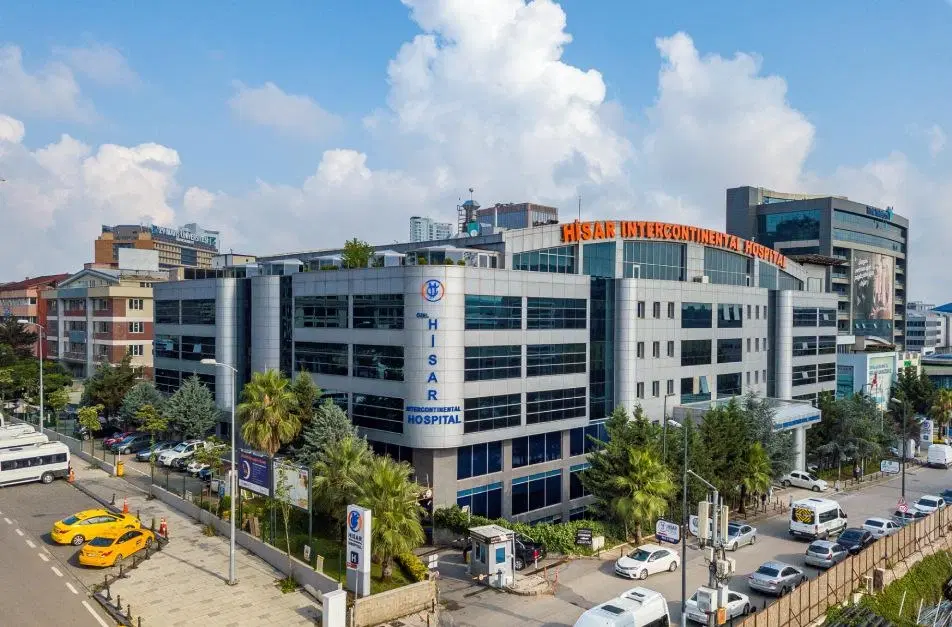

Hisar Intercontinental Hospital
Saray Mah. Siteyolu Cad. No:7, Umraniye, 34768, Istanbul, Turkey
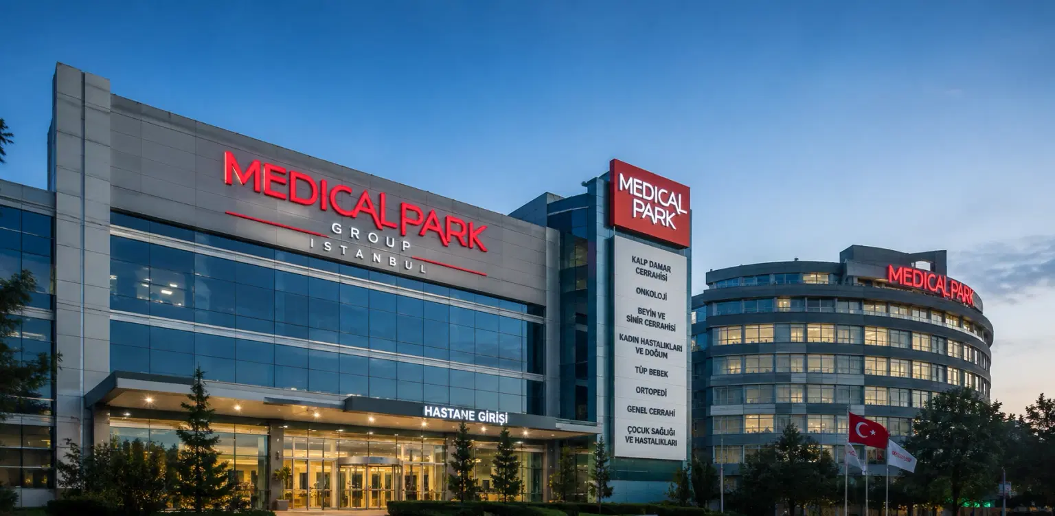
Medical Park Group, Istanbul
Fahrettin Kerim Gokay Cad. Tıbbiye Cd., Kadikoy, Istanbul, Turkey
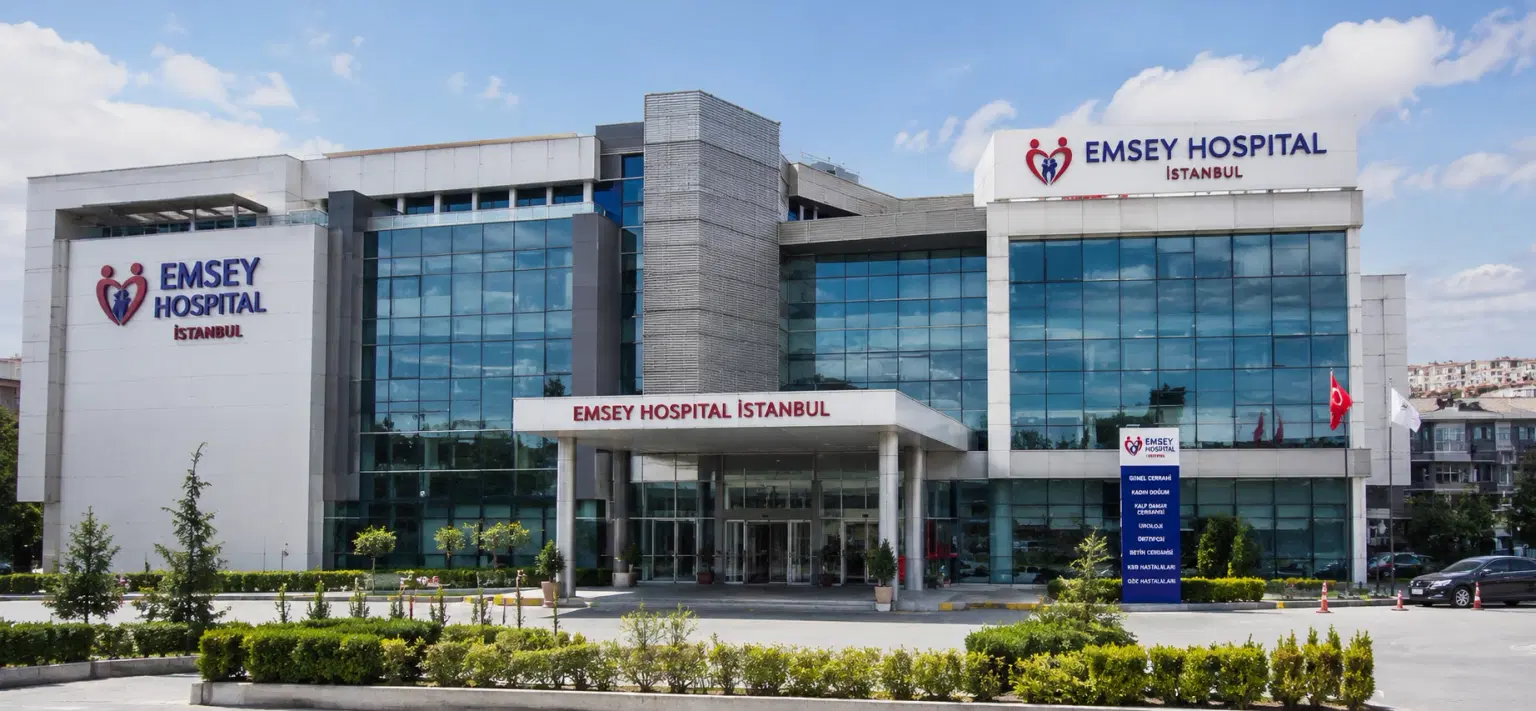
Emsey Hospital, Pendik, Istanbul
Çamlık, Selçuklu Cd. No:22, 34912 Pendik/İstanbul, Türkiye
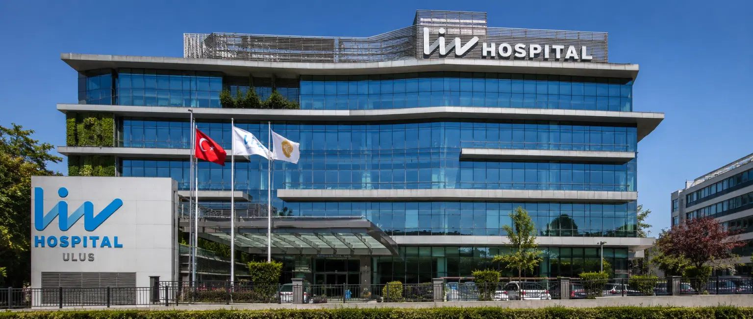
LIV Hospital, Istanbul
How DivinHeal Helps
We simplify your medical journey by providing comprehensive support and access to world-class healthcare.
Expert Specialist Matching
Connecting you with the world's top-rated medical experts.
Accredited Hospital Network
Access to JCI & NABH certified healthcare facilities.
Complete Travel Coordination
Hassle-free visa, stay, and local transport assistance.
24/7 Personal Care
Dedicated patient advisors supporting you at every step.
Journey Guidance
Full guidance from start to end of the patient treatment journey.
Expert Specialist Matching
Connecting you with the world's top-rated medical experts.
Everything you
need to know today
Browse through these common inquiries to better understand our patient-focused medical platform.
Yes, we work with a variety of insurance providers. Contact our team to verify your coverage.
Yes, we provide secure online consultations with experienced specialists.
Our care coordinators help match you with the most suitable specialist.
Absolutely. Your medical information is protected according to healthcare privacy standards.
Look at six things: accreditation (JCI or NABH), specialty depth, doctor credentials and experience, procedure-specific success rates, international patient support, and technology. DivinHeal's AI-driven matching evaluates every hospital in our accredited partner network on these dimensions and shortlists the best-fit options for your condition, budget, and country.
JCI (Joint Commission International) is the US-based global gold standard for hospital quality, recognised worldwide. NABH is India's national accreditation — accredited by ISQua, the same body that accredits JCI. Both signal independently verified safety and quality. Most of India's leading hospitals hold both.
Yes. All three welcome international patients through structured medical visa programs. India is the most established, treating patients from Africa, the Middle East, and South Asia at 60–80% lower cost. Thailand leads in cosmetic and dental care. The UAE is emerging in oncology and reproductive medicine.
Most patients save 50–80% on treatment costs. Heart bypass costs US $7,000–9,000 in India compared to $70,000–150,000 in the US. IVF costs $3,000–4,500 compared to $12,000–20,000 in the UK. Even after flights, visa, and accommodation, total savings remain 60–70%.
DivinHeal manages your entire non-medical journey: visa invitation letters, medical visa guidance, doctor appointments, teleconsultations, airport pickup, hospital-vetted accommodation for you and your attendant, language interpreters, local transport, cuisine preferences, and post-treatment follow-up — one dedicated coordinator from first enquiry to final follow-up.
You need a valid passport (6+ months validity), a medical visa (M-Visa for India — DivinHeal provides the hospital invitation letter), return flight tickets, recent medical reports and a doctor's referral, current prescription list, and proof of financial means. Any accompanying attendant needs their own passport and MX-Visa.
Still have more questions?
Book a call with our friendly team to learn how DivineHeal simplifies your healthcare journey.