



Deep Vein Thrombosis (DVT) Treatment / Thrombolysis
5K+ International Patients Treated
40+ Source Countries Served
500+ Accredited Partner Hospitals
98% Patient Satisfaction
80% Average Savings vs USA
10K+ Doctors
NABH, JCI Accredited Hospitals
Free Treatment Plan
Free Consultation with Doctor
5+ Destinations Covered
About Deep Vein Thrombosis (DVT) Treatment / Thrombolysis
Sources and Guidelines Referenced
The clinical protocols and recommendations in this guide are grounded in published clinical practice guidelines and major clinical trials: American College of Chest Physicians (CHEST) Guidelines on Antithrombotic Therapy for VTE Disease (2021 update); Society of Interventional Radiology (SIR) Quality Improvement Guidelines for the Performance of Lower Extremity Deep Vein Thrombolysis (2018); European Society of Cardiology (ESC) Guidelines on Diagnosis and Management of Acute Pulmonary Embolism (2019); National Institute for Health and Care Excellence (NICE) Guideline NG158 on Venous Thromboembolic Diseases (2020); The Acute Venous Thrombosis: Removal with Endovascular Therapy (ATTRACT) Trial (Vedantham et al., N Engl J Med, 2017); and The Catheter-Directed Venous Thrombolysis (CaVenT) Study (Enden et al., Lancet, 2012).
Deep Vein Thrombosis (DVT) Treatment / Thrombolysis: A Comprehensive Patient Guide
1. Definition and Medical Identity
Deep vein thrombosis treatment refers to the structured medical, endovascular, or surgical management of blood clots located in the body's deep venous system. The fundamental primary goals are stopping clot growth, preventing life-threatening pulmonary embolisms, and minimizing chronic venous valve damage known as post-thrombotic syndrome.
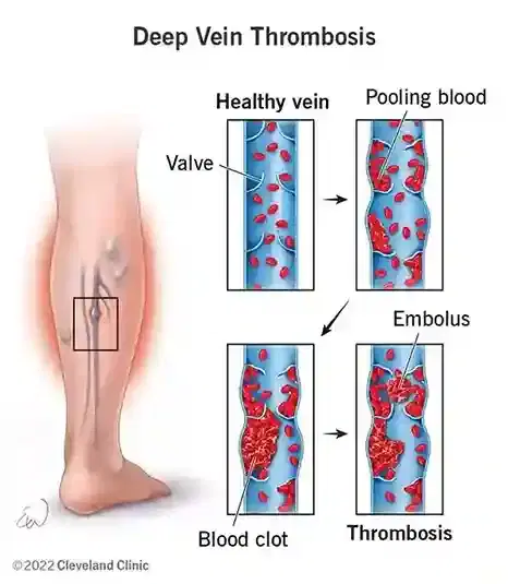
Deep vein thrombosis (DVT) occurs when blood solidifies within non-superficial veins, most commonly in the legs, pelvis, or upper extremities. The definitive treatment strategy depends on whether the clot is localized or extensive. Standard management involves anticoagulation therapy—medications colloquially called blood thinners—which prevent the body from forming additional blood clots while natural enzymatic mechanisms gradually break down existing fibrin structures. Advanced cases, particularly extensive upper-leg or pelvic blood clots, may require thrombolysis, a targeted procedure using thrombolytic agents (clot-dissolving drugs) to disintegrate the occlusion directly.
| Treatment Term | Medical Definition | Primary Clinical Mechanism |
|---|---|---|
| Anticoagulation | Systemic administration of drugs delaying blood coagulation pathways | Inhibits clotting factors (e.g., Factor Xa, thrombin) to prevent clot expansion |
| Catheter-Directed Thrombolysis (CDT) | Targeted delivery of plasminogen activators directly inside a blood clot via catheter | Converts plasminogen to plasmin to actively cleave fibrin strands within thrombus |
| Pharmacomechanical Thrombectomy (PMT) | Combination of localized drug delivery and mechanical clot maceration/aspiration | Mechanically breaks and vacuums dissolved thrombus for accelerated vein clearance |
| Surgical Venous Thrombectomy | Direct open surgical extraction of blood clot from an exposed vein | Manual surgical incision and removal of occlusive clot in acute limb-threatening cases |
2. The Underlying Condition or Need
Deep vein thrombosis develops when pathological blood clots obstruct deep venous channels, threatening systemic circulation and long-term limb health. Without timely therapy, thrombi can detach and migrate to the pulmonary arteries, or destroy vein valves and cause permanent leg pain, severe edema, and non-healing skin ulcers.
The deep veins return deoxygenated blood from the distal limbs back to the heart. When a thrombus forms, venous outflow is blocked, causing hydrostatic pressure to build up behind the occlusion. This acute venous hypertension leads to classic clinical presentation: unilateral leg swelling, calf tenderness, erythema (redness), and localized warmth. The condition is classically driven by Virchow's triad: hypercoagulability (increased blood clotting propensity), stasis (reduced blood flow velocity from immobility or obstruction), and endothelial injury (damage to the inner lining of blood vessels).
- Pulmonary Emembolism (PE): A life-threatening emergency where a portion of the DVT detaches, travels through the right side of the heart, and lodges in the pulmonary arterial bed, impairing gas exchange and causing strain on the right ventricle.
- Post-Thrombotic Syndrome (PTS): A chronic condition caused by venous valve destruction and persistent vein wall scarring, leading to permanent leg swelling, stasis dermatitis, hyperpigmentation, and painful leg ulcerations.
- Phlegmasia Cerulea Dolens: A massive, near-total occlusion of all venous outflow from a limb, causing severe cyanosis, ischemic tissue pain, arterial insufficiency, and potential gangrene if non-emergent intervention is delayed.
3. How the Treatment Works — Mechanism
DVT treatments operate by halting coagulation cascades or breaking down the cross-linked fibrin matrix within established blood clots. Anticoagulants prevent thrombus growth by suppressing enzymatic clotting factors, whereas thrombolytics convert inactive endogenous plasminogen into plasmin, an active enzyme that breaks down fibrin.
Standard medical therapy relies on anticoagulants, which do not actively dissolve established clots but suppress thrombin generation. By interrupting the clotting cascade, anticoagulants prevent the thrombus from enlarging, allowing the body's natural fibrinolytic system to slowly degrade the clot over weeks or months. Major anticoagulant drug classes include direct oral anticoagulants (DOACs) like apixaban and rivaroxaban, which inhibit Factor Xa; low-molecular-weight heparin (LMWH), which accelerates antithrombin activity; and vitamin K antagonists like warfarin, which inhibit hepatic synthesis of clotting factors II, VII, IX, and X.
For extensive, acute thrombi, direct mechanical or enzymatic dissolution is required. Catheter-directed thrombolysis (CDT) employs a specialized micro-catheter inserted into the clot under image guidance. The catheter delivers recombinant tissue plasminogen activator (rtPA, such as alteplase or reteplase). These agents bind to fibrin and convert localized plasminogen into plasmin. Plasmin hydrolyzes the fibrin network, rapidly liquefying the clot structure. In pharmacomechanical catheter-directed thrombolysis (PCDT), localized drug delivery is paired with mechanical devices—such as ultrasound-assisted catheters or rotational maceration tools—to break apart and aspirate the clot, reducing the required dose of thrombolytic drug and shortening procedure times.
4. Types and Variations
DVT treatment variations range from oral anticoagulant therapy to advanced image-guided endovascular interventions and surgical options. Clinicians select protocols based on anatomical location, duration of symptoms, bleeding risk profile, and overall clinical severity.
Treatment selection follows established clinical algorithms, such as those published by the American College of Chest Physicians (CHEST 2021). Distal (calf) deep vein thrombosis without severe symptoms is often managed with initial surveillance or short-term oral anticoagulation. Proximal DVT (popliteal, femoral, or iliac veins) requires immediate therapeutic anticoagulation. Catheter-directed interventions are selectively used for acute iliofemoral DVT (clots extending into the groin and pelvic veins) presenting within 14 days of symptom onset in low-bleeding-risk candidates.
| Treatment Variation | Invasiveness | Typical Setting | Clinical Indications | Primary Trade-Offs |
|---|---|---|---|---|
| Direct Oral Anticoagulants (DOACs) | Non-invasive (Oral) | Outpatient | First-line therapy for distal and routine proximal DVT | Low bleeding risk; slow natural clot breakdown |
| Catheter-Directed Thrombolysis (CDT) | Minimally Invasive (Endovascular) | Inpatient / ICU | Acute, extensive iliofemoral DVT (<14 days duration) | Rapid clot clearance; elevated major bleeding risk |
| Pharmacomechanical Thrombectomy (PCDT) | Minimally Invasive (Endovascular) | Inpatient / Angio Suite | Extensive DVT where lower drug exposure is preferred | Shorter treatment duration; requires specialized endovascular tools |
| Ultrasound-Assisted Thrombolysis (USAT) | Minimally Invasive (Endovascular) | Inpatient / ICU | Severe proximal DVT; combined mechanical-lytic therapy | High clot penetration; requires intensive monitoring |
| Surgical Venous Thrombectomy | Invasive Surgical | Operating Room | Phlegmasia cerulea dolens when thrombolysis is contraindicated | Immediate pressure relief; surgical morbidity and high re-thrombosis risk |
5. Who the Treatment Is For — Indications
DVT treatment is indicated for all confirmed cases of acute deep vein thrombosis to eliminate the immediate risk of pulmonary embolism. Catheter-directed thrombolysis is reserved for candidates with acute, extensive iliofemoral thrombosis, acceptable bleeding risks, and a functional recovery potential.
Diagnostic evaluation begins with a structured clinical pre-test probability assessment (such as the Wells Score). If DVT is clinically suspected, diagnostic imaging is performed:
- Compression Duplex Ultrasonography: The primary non-invasive diagnostic standard. Inability to compress the vein wall under ultrasound probe pressure confirms the presence of an intraluminal thrombus.
- D-Dimer Testing: A blood test measuring fibrin degradation products, used primarily to rule out venous thromboembolism in low-probability patients.
- Computed Tomography (CT) Venography or Magnetic Resonance (MR) Venography: Advanced cross-sectional imaging used to define the upper extent of pelvic, iliac, or inferior vena cava (IVC) blood clots.
Specific indications for advanced endovascular thrombolysis (SIR Guidelines 2018) include: acute symptomatic iliofemoral DVT with symptoms lasting fewer than 14 days, low predicted risk of bleeding complications, good baseline functional capacity, a life expectancy greater than one year, and severe swelling or limb-threatening vascular compromise.
6. Who the Treatment Is Not For — Contraindications
Contraindications to standard DVT therapy depend on the specific intervention, primarily driven by absolute and relative bleeding risks. While anticoagulation is rarely withheld entirely, catheter-directed thrombolysis carries strict safety boundaries due to the risk of major hemorrhage.
Absolute contraindications to catheter-directed thrombolysis (CHEST 2021; SIR 2018) include active internal bleeding, recent intracranial hemorrhage, structural cerebrovascular lesions, active intracranial or spinal neoplasm, ischemic stroke within the preceding 3 months, major surgery or severe neurotrauma within the past 2 to 4 weeks, and absolute bleeding diathesis.
| Contraindication Category | Clinical Scenario | Therapeutic Adjustment / Action |
|---|---|---|
| Absolute (Thrombolysis) | Active internal bleeding or recent hemorrhagic stroke | Cancel thrombolysis; evaluate for temporary Inferior Vena Cava (IVC) filter |
| Absolute (Thrombolysis) | Recent neurosurgery or severe head trauma (<4 weeks) | Avoid thrombolytic drugs; rely on therapeutic anticoagulation or mechanical options |
| Relative (Thrombolysis) | Severe uncontrolled hypertension (BP > 180/110 mmHg) | Pharmacologically lower blood pressure prior to considering endovascular catheter placement |
| Relative (Anticoagulation) | Severe thrombocytopenia (platelet count < 50,000/µL) | Reduce drug dosage or select short-acting agents with continuous laboratory monitoring |
| Absolute (DOACs) | Severe renal impairment (CrCl < 15–30 mL/min) or Antiphospholipid Syndrome | Switch from DOACs to unfractionated heparin, LMWH, or Vitamin K Antagonists (Warfarin) |
7. Alternatives and Clinical Comparison
Alternatives to catheter-directed thrombolysis include standard systemic anticoagulation alone, inferior vena cava filters, and conservative mechanical compression measures. Clinical trial evidence guides the selection between aggressive clot removal and non-invasive medical management.
The landmark ATTRACT Trial (Vedantham et al., N Engl J Med, 2017) evaluated 692 patients with acute proximal DVT, comparing anticoagulation alone to pharmacomechanical catheter-directed thrombolysis plus anticoagulation. While PCDT did not reduce overall 2-year post-thrombotic syndrome incidence, it significantly reduced early acute leg pain and swelling, as well as moderate-to-severe PTS rates in the subgroup with true iliofemoral DVT. Similarly, the CaVenT Trial (Enden et al., Lancet, 2012) demonstrated a significant absolute risk reduction in PTS at 2 and 5 years following catheter-directed thrombolysis.
| Treatment Option | Mechanism | Primary Advantage | Primary Disadvantage / Risk | Best Suited For |
|---|---|---|---|---|
| Systemic Anticoagulation (DOACs) | Inhibits coagulation enzymes | Oral dosing; low major bleeding risk | Slow natural clot resolution; potential long-term valve scarring | Routine distal and femoropopliteal DVT |
| Catheter-Directed Thrombolysis (CDT) | Targeted intra-clot fibrinolysis | Rapid thrombus dissolution; preserves venous valves | Higher major bleeding risk; requires hospital/ICU admission | 选Acute iliofemoral DVT with low bleeding risk |
| Inferior Vena Cava (IVC) Filter | Mechanical capture of dislodged emboli | Prevents pulmonary embolism without raising bleeding risk | Does not treat underlying clot; risk of filter thrombosis or vessel penetration | Patients with acute DVT and absolute contraindications to anticoagulation |
| Graduated Compression Alone | External mechanical compression | Non-invasive; zero systemic risk | Does not prevent clot growth or pulmonary embolism on its own | Adjunct management; isolated low-risk distal DVT without expansion criteria |
8. Pre-Treatment Phase
The pre-treatment phase involves immediate patient stabilization, rapid diagnostic verification, bleeding risk evaluation, and pre-procedural optimization. For endovascular interventions, precise vascular mapping and laboratory screening are completed before entering the interventional suite.
Initial history-taking focuses on symptom duration, prior thrombotic events, active bleeding risks, personal or family history of hypercoagulability, active malignancies, and recent surgical procedures. Laboratory testing must include a complete blood count (to check baseline hemoglobin and platelet levels), coagulation panels (PT/INR, aPTT), baseline renal panel (serum creatinine and eGFR for contrast planning), and liver function tests.
- Medication Adjustment: Non-essential antiplatelet drugs (such as aspirin or clopidogrel) or non-steroidal anti-inflammatory drugs (NSAIDs) are evaluated and paused if catheter interventions are planned. If the patient is already taking anticoagulants, specific bridging protocols are instituted.
- Informed Consent: The patient is counseled on the procedure's goals, expected symptom relief, potential major bleeding risks (including a rare risk of hemorrhagic stroke), and the potential need for balloon angioplasty or venous stenting to correct underlying vein compressions.
- Procedural Preparation: The patient remains nil per os (NPO) for 6 hours prior to endovascular interventions. Intravenous access is established, and baseline limb circumferences are measured to monitor swelling reduction.
9. The Procedure — Step-by-Step Clinical Detail
Catheter-directed thrombolysis is a multi-step endovascular procedure performed in an interventional radiology or vascular catheterization laboratory. Image guidance is used throughout to accurately target and dissolve venous blood clots.
The procedural workflow follows these standardized steps:
- Vascular Access: The patient is positioned prone or supine on the fluoroscopy table. Under real-time ultrasound guidance, the interventional physician accesses a clear venous segment below the clot—typically the popliteal vein, posterior tibial vein, or small saphenous vein—using a micro-puncture kit and local anesthesia with conscious sedation.
- Diagnostic Venography: A vascular sheath is placed, and contrast catheter venography is performed under fluoroscopy to precisely visualize the lower and upper limits of the thrombus.
- Catheter Positioning: A specialized multi-sidehole infusion catheter is advanced over a guidewire directly through the body of the clot.
- Thrombolytic Drug Infusion: Recombinant tissue plasminogen activator (rtPA) is initiated at a low continuous rate (e.g., 0.5–1.0 mg/hour per catheter). Sub-therapeutic unfractionated heparin is infused concurrently through the sheath to prevent pericatheter thrombosis.
- Pharmacomechanical Enhancement (Optional): If PCDT is utilized, dedicated mechanical thrombectomy catheters or ultrasound-assisted devices are deployed to physically disrupt or vacuum dissolved clot fragments.
- Serial Venographic Checks: The patient is transferred to an intensive care or step-down unit while infusion continues over 12 to 24 hours. The patient returns to the suite for repeat venography to assess progress.
- Angioplasty and Stenting: Once the clot is cleared, underlying anatomical narrowing—such as iliac vein compression (May-Thurner syndrome)—is routinely identified. The clinician performs balloon angioplasty and deploys a dedicated, wide-diameter flexible venous stent to maintain long-term vessel open rate (patency).
- Hemostasis: The catheter is removed, and manual compression is applied to the puncture site until complete hemostasis is achieved.
10. Immediate Post-Procedure Period
The immediate 24 to 48 hours following standard treatment or endovascular thrombolysis requires intensive clinical monitoring to detect bleeding, verify access-site stability, and confirm re-established venous blood flow. Early mobilization is encouraged as soon as access site stability is confirmed.
Patients undergoing catheter-directed thrombolysis are monitored closely in a high-acuity setting during active drug infusion. Vital signs, neurological checks (to monitor for severe intracranial bleeding), and catheter site inspections are conducted every 1 to 2 hours. Fibrinogen blood levels, hemoglobin, and platelet counts are drawn every 6 to 8 hours; if plasma fibrinogen drops below critical thresholds (e.g., < 100–150 mg/dL), the thrombolytic infusion rate is reduced or paused to prevent bleeding complications.
Following catheter sheath removal, absolute bed rest with the limb extended is maintained for 4 to 6 hours. Once puncture site hemostasis is verified, gentle walking is reinitiated. Full therapeutic anticoagulation is re-established, and medical-grade graduated compression stockings (30–40 mmHg or 20–30 mmHg) are fitted to aid the calf muscle pump and reduce localized edema.
11. Recovery — Short and Long Term
Recovery following DVT treatment spans several phases, from initial swelling reduction in the first two weeks to long-term venous health management over 3 to 12 months. Strict adherence to medical therapy and lifestyle adjustments supports optimal recovery.
During the first 1 to 2 weeks, acute leg pain typically resolves, and leg circumference returns toward baseline. Patients are encouraged to ambulate multiple times daily while avoiding prolonged periods of standing or sitting. Limb elevation above heart level during rest helps minimize edema accumulation.
- 1 to 4 Weeks: Continuation of oral anticoagulation (DOACs). Daily walking goals are gradually increased. Patients can return to desk work within several days and light physical activity within 2 to 3 weeks. High-impact sports or contact activities are avoided while on therapeutic anticoagulation.
- 1 to 3 Months: First follow-up visit with duplex ultrasonography to assess vein patency, venous valve competence, and stent stability (if placed). Anticoagulation duration is re-evaluated based on whether the original DVT was provoked (e.g., surgery, trauma) or unprovoked.
- 6 to 12 Months: Long-term outcome assessment for signs of post-thrombotic syndrome using standardized clinical tools like the Villalta Scale. Decision-making on extending anticoagulation indefinitely based on persistent risk factors or elevated D-dimer levels following brief treatment pauses.
12. Risks, Side Effects, and Complications
DVT treatments carry inherent risks, ranging from minor bruising to major, life-threatening hemorrhagic events. Medical strategies balance the benefit of clot clearance and stroke prevention against bleeding risks.
Major bleeding represents the most significant adverse risk associated with both oral anticoagulation and thrombolysis. According to data from the ATTRACT trial (Vedantham et al., 2017), major bleeding occurs in approximately 1.7% of patients receiving standard anticoagulation and up to 4.7% of patients undergoing pharmacomechanical thrombolysis. Intracranial hemorrhage—the most catastrophic complication—occurs in less than 1% of CDT cases but requires immediate cessation of therapy and emergency intervention.
| Risk Category | Complication / Side Effect | Incidence Rate | Clinical Management Strategy |
|---|---|---|---|
| Common / Mild | Access-site hematoma or minor ecchymosis | 10% – 20% | Local manual pressure, cold compresses, clinical monitoring |
| Common / Mild | Minor mucosal bleeding (epistaxis, gingival) | 5% – 15% | Reassurance, patient education, temporary medication review |
| Uncommon / Moderate | Access-site major vascular injury or pseudoaneurysm | 1% – 3% | Ultrasound-guided compression or thrombin injection |
| Uncommon / Moderate | Contrast-Induced Nephropathy (CIN) | 1% – 3% | Periprocedural intravenous hydration; contrast volume minimization |
| Rare / Serious | Major gastrointestinal or retroperitoneal hemorrhage | 1% – 4% | Discontinue anticoagulants/lytics; administer reversal agents and blood products |
| Rare / Serious | Symptomatic Intracranial Hemorrhage (ICH) | 0.5% – 1.0% | Immediate therapy withdrawal, emergency non-contrast head CT, factor replacement |
| Rare / Serious | Pulmonary Embolism during mechanical manipulation | < 1% | Aspiration thrombectomy, emergency respiratory and hemodynamic support |
13. Lifestyle and Behavioural Considerations
Behavioral adjustments play a primary supportive role in recovering from DVT and preventing recurrent venous thromboembolism. Patients can make evidence-based lifestyle adaptations during and after medical therapy to lower recurrence risks.
Key recommendations supported by clinical guidelines include:
- Active Mobilization: Regular walking stimulates the calf muscle pump, forcing venous blood upward and countering stasis. Patients should avoid sitting or standing still for longer than 60 minutes at a time.
- Graduated Compression Garments: Wearing medical-grade compression stockings provides targeted external counter-pressure, preventing blood pooling and alleviating symptoms of chronic venous insufficiency.
- Hydration and Weight Management: Maintaining appropriate fluid intake prevents hemoconcentration, while achieving a healthy body weight reduces intra-abdominal pressure that can impede venous return from the lower limbs.
- Long-Distance Travel Adaptations: During long flights or automobile trips (>4 hours), patients should perform frequent calf-isometric exercises, flex their ankles, wear compression garments, and walk around every 1 to 2 hours.
14. How Outcomes Are Measured
Clinical success following DVT treatment is evaluated through symptom resolution, ultrasound findings, patency rates, and the prevention of chronic complications like post-thrombotic syndrome.
Short-term success is defined as rapid pain relief, a measurable decrease in limb circumference, and restoring vein open rate (patency), as confirmed by follow-up compression duplex ultrasound within 30 days. Complete thrombus clearance (greater than 50% to 90% lumen restoration) is the primary procedural goal of catheter-directed thrombolysis.
Long-term clinical efficacy is measured using the validated Villalta Scale, which quantifies five patient-reported symptoms (pain, cramps, heaviness, paresthesia, pruritus) and six clinical signs (edema, skin induration, hyperpigmentation, venous ectasia, redness, pain on calf compression). A Villalta score under 5 indicates the absence of post-thrombotic syndrome, whereas a score of 15 or higher (or the presence of a venous ulcer) indicates severe PTS. Long-term studies demonstrate that successful early clot removal via CDT reduces severe PTS incidence over a 2- to 5-year follow-up window (Enden et al., 2012).
15. Recent Advances and Current Standard of Care
The standard of care for deep vein thrombosis has evolved significantly over the past decade, driven by novel direct oral anticoagulants, catheter technologies, and refined treatment selection guidelines.
Major advances transforming DVT care include:
- Direct Oral Anticoagulants (DOACs): Factor Xa inhibitors (apixaban, rivaroxaban) and direct thrombin inhibitors (dabigatran) have largely replaced traditional vitamin K antagonists (warfarin). DOACs offer predictable pharmacokinetics, eliminate the need for routine laboratory monitoring (INR testing), and carry a lower risk of intracranial bleeding.
- Single-Session Pharmacomechanical Thrombectomy: Next-generation mechanical thrombectomy systems utilize large-bore aspiration catheters or acoustic ultrasound energy to evacuate blood clots in a single interventional session. This eliminates the need for prolonged overnight ICU thrombolytic infusions, lowering major bleeding risks.
- Dedicated Venous Stents: The development of self-expanding, highly flexible nickel-titanium (nitinol) stents specifically designed for the high-tonus venous system (such as the iliac veins) has improved long-term vessel patency and reduced stent structural failure compared to older arterial stents.
16. Common Myths and Misconceptions
Misunderstandings regarding DVT treatment can cause unnecessary anxiety or lead to non-adherence with essential medical therapies. Clarifying these clinical concepts helps establish realistic treatment expectations.
Myth: Blood thinners immediately dissolve existing blood clots.
Reality: Anticoagulants do not actively dissolve established blood clots; they stop the clot from growing and prevent new clots from forming, allowing the body's natural enzymes to slowly break down the thrombus over time.
Myth: Bed rest is mandatory after being diagnosed with a leg DVT.
Reality: Early ambulation combined with effective anticoagulation is safe and recommended over prolonged bed rest, as walking helps improve venous return without increasing pulmonary embolism risk (CHEST 2021 Guidelines).
Myth: Once DVT pain vanishes, anticoagulation can be stopped.
Reality: Prematurely stopping blood thinners greatly increases the risk of recurrent thrombosis. Medication duration is determined by clinical guidelines based on thrombotic risk profiles, regardless of early symptom resolution.
Myth: Catheter-directed thrombolysis is necessary for all leg blood clots.
Reality: Catheter-directed thrombolysis is selectively reserved for severe, extensive iliofemoral DVT in patients with low bleeding risks, whereas standard anticoagulation remains the primary recommendation for most proximal and distal clots (ATTRACT Trial, 2017).
Myth: As long as you take blood thinners, you will never develop another DVT.
Reality: While anticoagulants lower recurrent thrombosis risk by over 80–90%, breakthrough blood clots can still occur due to underlying anatomical compressions, active cancer, or medication non-compliance.
Myth: Leg swelling after DVT means the clot has returned.
Reality: Persistent or recurrent swelling is frequently caused by post-thrombotic syndrome due to prior vein valve damage, rather than a new active blood clot, though ultrasound evaluation is required to differentiate the two.
17. Frequently Asked Questions
What is the difference between anticoagulation and thrombolysis?
Anticoagulants are systemic oral or intravenous medications that stop new clots from forming and prevent existing clots from growing, allowing your body to process the clot over time. Thrombolysis is an endovascular procedure where clot-dissolving drugs are delivered directly into the thrombus via a small catheter to rapidly disintegrate the clot structure within hours.
How long will I need to take blood thinners for a DVT?
The duration of anticoagulation therapy depends on whether the DVT was provoked by a clear transient risk factor, such as recent surgery, or was unprovoked. First-episode provoked DVTs are typically treated for 3 to 6 months. Unprovoked DVTs, recurrent clots, or cases with ongoing underlying risk factors often require extended or lifelong therapy, guided by periodic risk reassessments.
Is catheter-directed thrombolysis painful?
The catheter placement process is performed under local anesthesia and conscious sedation, minimizing discomfort. You may feel slight pressure at the vein entry site (usually behind the knee or in the groin). During thrombolytic infusion or mechanical clot retrieval, mild aching or pressure in the leg can occur, which is managed with intravenous analgesics.
When can I safely resume normal exercise after DVT treatment?
Gentle walking is encouraged immediately once your condition is stabilized on anticoagulation. Light aerobic activity can typically resume within 1 to 2 weeks as leg pain and swelling subside. High-impact sports, contact athletics, or heavy weightlifting should be deferred until clearing the initial recovery phase, especially while taking high-dose blood thinners that increase bleeding risks.
What is May-Thurner syndrome and how does it relate to DVT?
May-Thurner syndrome is an anatomical variation where the left common iliac vein is compressed by the overlying right common iliac artery against the spine. This chronic mechanical compression stanches venous outflow, significantly increasing the risk of left leg deep vein thrombosis. It is frequently identified and treated with balloon angioplasty and stenting following clot removal.
How do compression stockings help treat DVT?
Medical-grade graduated compression stockings exert controlled, descending pressure from the ankle upward. This external compression reduces vein diameter, speeds up deep venous blood flow, prevents blood pooling, restores valve function, and alleviates leg pain and swelling during daily activities.
What are the warning signs of a dangerous bleeding complication?
Signs of major internal bleeding while taking anticoagants or receiving thrombolysis include unusual severe headaches, sudden dizziness or confusion, coughing up blood, vomiting dark material resembling coffee grounds, passage of black or bright red stools, unexpected deep bruising, or uncontrolled bleeding from small cuts. Immediate medical evaluation is required if these occur.
Can a blood clot in the leg travel to the brain and cause a stroke?
Under normal anatomical conditions, a DVT in the venous system travels through the right side of the heart to the lungs, causing a pulmonary embolism, rather than reaching the brain. However, if a cardiac defect like a patent foramen ovale (PFO) is present, a venous clot can cross into the arterial circulation and travel to the brain, causing a paradoxical ischemic stroke.
How long does leg swelling last after DVT treatment?
Acute edema usually decreases significantly within 2 to 4 weeks of starting treatment. However, mild to moderate leg swelling may persist for several months if the deep vein valves suffered structural damage. Wearing compression stockings and elevating the leg during rest helps manage ongoing fluid accumulation.
What is an IVC filter and when is it necessary?
An inferior vena cava (IVC) filter is a small, basket-like metal device placed in the main abdominal vein to trap large blood clots before they reach the lungs. It is reserved for patients with acute, high-risk DVT who have absolute contraindications to blood thinners (such as active severe internal bleeding) or those who develop recurrent pulmonary emboli despite therapeutic anticoagulation.
Will I need a repeat ultrasound during my recovery?
Yes, follow-up compression duplex ultrasound scans are routinely scheduled at 1 to 3 months and potentially at 6 months. These scans verify that the blood clot has stabilized or dissolved, assess long-term blood vessel patency, check for residual vein narrowing, and evaluate whether implanted venous stents remain fully open.
How soon can I travel on a commercial airplane after a DVT?
Short air travel may be permitted within 1 to 2 weeks once therapeutic anticoagulation is established and acute symptoms resolve. For long-distance flights (>4 hours), patients should defer travel for at least 4 weeks if possible, wear compression stockings, maintain hydration, and perform frequent ankle flexing and walking during the flight.
Booking With DIVINHEAL
Get a free consultation to understand your treatment options
Cost Calculator
I know my treatment - show me cost from 3 hospitals
Plan My Journey
Tell us your condition and budget - our AI matches the right destination, hospital and doctor and visa pathway
Recommended Article

Booking With DIVINHEAL
Get a free consultation to understand your treatment options
Cost Calculator
I know my treatment - show me cost from 3 hospitals
Plan My Journey
Tell us your condition and budget - our AI matches the right destination, hospital and doctor and visa pathway
Recommended Article
Meet Our Medical Specialists
Hospitals
NABH & JCI Accredited Hospitals in India,Turkey, Thailand & UAE.
How DivinHeal Helps
We simplify your medical journey by providing comprehensive support and access to world-class healthcare.
Expert Specialist Matching
Connecting you with the world's top-rated medical experts.
Accredited Hospital Network
Access to JCI & NABH certified healthcare facilities.
Complete Travel Coordination
Hassle-free visa, stay, and local transport assistance.
24/7 Personal Care
Dedicated patient advisors supporting you at every step.
Journey Guidance
Full guidance from start to end of the patient treatment journey.
Expert Specialist Matching
Connecting you with the world's top-rated medical experts.
Everything you
need to know today
Browse through these common inquiries to better understand our patient-focused medical platform.
Yes, we work with a variety of insurance providers. Contact our team to verify your coverage.
Yes, we provide secure online consultations with experienced specialists.
Our care coordinators help match you with the most suitable specialist.
Absolutely. Your medical information is protected according to healthcare privacy standards.
Look at six things: accreditation (JCI or NABH), specialty depth, doctor credentials and experience, procedure-specific success rates, international patient support, and technology. DivinHeal's AI-driven matching evaluates every hospital in our accredited partner network on these dimensions and shortlists the best-fit options for your condition, budget, and country.
JCI (Joint Commission International) is the US-based global gold standard for hospital quality, recognised worldwide. NABH is India's national accreditation — accredited by ISQua, the same body that accredits JCI. Both signal independently verified safety and quality. Most of India's leading hospitals hold both.
Yes. All three welcome international patients through structured medical visa programs. India is the most established, treating patients from Africa, the Middle East, and South Asia at 60–80% lower cost. Thailand leads in cosmetic and dental care. The UAE is emerging in oncology and reproductive medicine.
Most patients save 50–80% on treatment costs. Heart bypass costs US $7,000–9,000 in India compared to $70,000–150,000 in the US. IVF costs $3,000–4,500 compared to $12,000–20,000 in the UK. Even after flights, visa, and accommodation, total savings remain 60–70%.
DivinHeal manages your entire non-medical journey: visa invitation letters, medical visa guidance, doctor appointments, teleconsultations, airport pickup, hospital-vetted accommodation for you and your attendant, language interpreters, local transport, cuisine preferences, and post-treatment follow-up — one dedicated coordinator from first enquiry to final follow-up.
You need a valid passport (6+ months validity), a medical visa (M-Visa for India — DivinHeal provides the hospital invitation letter), return flight tickets, recent medical reports and a doctor's referral, current prescription list, and proof of financial means. Any accompanying attendant needs their own passport and MX-Visa.
Still have more questions?
Book a call with our friendly team to learn how DivineHeal simplifies your healthcare journey.