



Doppler Ultrasound
5K+ International Patients Treated
40+ Source Countries Served
500+ Accredited Partner Hospitals
98% Patient Satisfaction
80% Average Savings vs USA
10K+ Doctors
NABH, JCI Accredited Hospitals
Free Treatment Plan
Free Consultation with Doctor
5+ Destinations Covered
About Doppler Ultrasound
Sources and Guidelines Referenced
This clinical guide is grounded in international consensus standards and evidence-based practice guidelines published by leading medical authorities, including: American Institute of Ultrasound in Medicine (AIUM Practice Parameter for Vascular Examinations, 2023); American College of Radiology (ACR Appropriateness Criteria for Suspected Deep Vein Thrombosis and Vascular Disease, 2022); Society for Vascular Ultrasound (SVU Professional Performance Standards, 2021); European Society of Cardiology (ESC Guidelines on Diagnosis and Treatment of Peripheral Arterial Diseases, 2021); American College of Obstetricians and Gynecologists (ACOG Practice Bulletin No. 204: Fetal Growth Restriction, 2020); World Health Organization (WHO Manual of Diagnostic Ultrasound, 2021); and Society of Radiologists in Ultrasound (SRU Consensus Guidelines on Carotid Artery Stenosis, 2020).
Doppler Ultrasound: A Comprehensive Patient Guide
1. Definition and Medical Identity
A Doppler ultrasound is a non-invasive diagnostic medical imaging examination that uses high-frequency sound waves to measure and visualize blood flow through arterial and venous blood vessels. Categorized under non-invasive vascular diagnostic radiology, its primary clinical goal is to assess vascular patency, detect blockages or clots, and quantify blood velocity.
Also known as vascular ultrasonography, duplex ultrasound, or Doppler sonography, this diagnostic technique combines traditional anatomical ultrasound with blood flow motion detection. Standard non-Doppler ultrasound produces detailed static structural images of internal organs and tissue layers by calculating the time it takes for acoustic echo waves to bounce back from soft tissues. In contrast, Doppler ultrasound specifically targets moving targets—primarily erythrocytes (red blood cells)—within blood vessels.
By capturing the physical shift in sound frequencies caused by moving blood, the system converts acoustic data into audible heart sound signals, visual spectral velocity graphs, and dynamic multi-color overlay images. Diagnostic radiologists and vascular specialists rely on this capability to assess blood circulation in major anatomical regions, including the head and neck, heart, abdomen, upper extremities, lower extremities, and fetal-placental circulation.
2. The Underlying Condition or Need
Doppler ultrasound addresses vascular conditions caused by impaired or obstructed blood flow, including arterial narrowing, blood clots, valve failure, and circulatory defects. Clinicians utilize this test when patients present with swelling, pain, numbness, or abnormal pulses to prevent severe complications like stroke, tissue death, or pulmonary embolism.
Vascular diseases represent a major global cause of disability and mortality. When the structural integrity or inner diameter of a blood vessel is compromised, local tissue perfusion decreases, leading to tissue ischemia, swelling, or systemic metabolic stress. Pathological mechanisms evaluated by Doppler ultrasound fall into three main functional categories:
- Luminal Obstruction: Intravascular blockages caused by acute blood clot formation—such as deep vein thrombosis (DVT) in the legs—or chronic atherosclerotic plaque accumulation, seen in carotid artery stenosis and peripheral artery disease (PAD).
- Valvular Incompetence: Failure of tiny, one-way venous valves inside deep or superficial leg veins, allowing blood to flow backward under gravity (venous reflux). This pathology causes chronic venous insufficiency (CVI), leg swelling, skin discoloration, and varicose veins.
- Hemodynamic and Structural Alterations: Abnormal blood vessel enlargement, such as abdominal aortic aneurysms, microvascular damage from renal disease, arterial dissection, or congenital vascular malformations.
Without accurate diagnostic visualization, untreated deep vein thrombosis can dislodge and travel to the lungs, causing a life-threatening pulmonary embolism (PE). Similarly, undetected carotid plaque buildup can break loose and block cerebral arteries, causing an ischemic stroke (ACR Appropriateness Criteria 2022). Doppler ultrasound provides the non-invasive clinical evaluation needed to identify these conditions early and guide targeted medical, endovascular, or surgical treatment.
3. How the Treatment Works — Mechanism
Doppler ultrasound functions using the physical principle of the Doppler shift, where sound waves change frequency when bouncing off moving objects like red blood cells. A hand-held probe sends acoustic waves into tissue; the frequency shift of returning waves calculates blood flow speed, direction, and turbulence in real time.
The foundation of this technology is the Doppler effect, first described mathematically by physicist Christian Doppler in 1842. When a diagnostic ultrasound probe (transducer) presses against the skin, it emits high-frequency, non-audible acoustic pulses—typically between 2.0 and 15.0 megahertz (MHz)—into underlying anatomical tissues.
When these sound waves hit stationary structures like muscle or liver tissue, they bounce back to the transducer at the exact same acoustic frequency. However, when sound waves strike moving red blood cells within an artery or vein, the reflected frequency changes:
- Positive Doppler Shift: Blood cells moving toward the transducer compress the reflected sound waves, returning a higher frequency than emitted.
- Negative Doppler Shift: Blood cells moving away from the transducer stretch the reflected sound waves, returning a lower frequency than emitted.
The ultrasound computer measures this frequency shift ($ \Delta f$) using the mathematical formula:
$$\Delta f = \frac{2 \cdot f_0 \cdot v \cdot \cos(\theta)}{c}$$
Where $f_0$ represents the emitted frequency, $v$ is blood flow velocity, $\theta$ is the angle of beam insonation, and $c$ is sound propagation speed in human tissue (approximately 1,540 meters per second). Because the cosine of 90 degrees equals zero, keeping the probe angled between 45 and 60 degrees relative to the blood vessel is essential for accurate velocity measurements (AIUM Guidelines 2023).
The ultrasound machine converts calculated velocity signals into graphical waveform tracing (spectral Doppler) or color-coded visual displays (color Doppler). Red typically represents blood flowing toward the probe, while blue represents flow moving away. Brightness indicates relative velocity or signal intensity.
4. Types and Variations
Doppler ultrasound encompasses several technical modalities, including Continuous Wave Doppler, Pulsed Wave Doppler, Color Doppler, Power Doppler, and Duplex Ultrasound. Clinicians select specific variations based on target vessel depth, flow velocity requirements, structural imaging needs, and whether low-velocity microvascular detection is necessary.
Ultrasound platforms incorporate different Doppler modes depending on clinical goals. Modern diagnostic equipment combines several modalities into a single evaluation.
| Doppler Modality | Acoustic Mechanism | Primary Clinical Use | Key Technical Advantage | Main Limitation |
|---|---|---|---|---|
| Continuous Wave (CW) Doppler | Uses separate crystals to continuously send and receive sound signals. | Bedside ankle-brachial index testing, fetal heart rate monitoring. | Accurately measures extremely high blood velocities without signal distortion. | Lacks depth resolution; cannot pinpoint exact depth of blood flow. |
| Pulsed Wave (PW) Doppler | Sends short sound pulses and waits for echo returns from specific depths. | Measuring specific peak blood speeds in carotid or renal arteries. | Provides precise depth targeting and quantitative velocity graphs. | Subject to signal distortion (aliasing) at very high blood flow speeds. |
| Color Doppler | Converts frequency shifts into color patterns over standard tissue images. | Mapping leg vein flow, locating turbulent arterial narrowing. | Rapidly surveys broad vascular pathways and visualizes direction. | Qualitative only; does not provide exact numerical velocity measurements. |
| Power Doppler | Displays total strength (amplitude) of reflected acoustic signals. | Evaluating blood flow in kidneys, tumors, or twisted testicles. | Five to ten times more sensitive for detecting slow microvascular flow. | Does not show blood flow direction or measure numerical speeds. |
| Duplex / Triplex Ultrasound | Combines B-mode tissue imaging, Color Doppler, and Pulsed Wave spectral tracing. | Gold-standard comprehensive arterial and venous examinations. | Simultaneously evaluates anatomical structure and exact blood velocity. | Requires advanced sonographer technical expertise and equipment. |
Selection among these modalities depends on diagnostic intent. For example, a vascular sonographer assessing peripheral artery disease uses B-mode structural imaging to locate arterial wall plaque, Color Doppler to highlight narrow areas of turbulent flow, and Pulsed Wave Doppler to measure exact peak systolic velocity (PSV) across narrowed segments (SVU Performance Standards 2021).
5. Who the Treatment Is For — Indications
Doppler ultrasound is indicated for individuals presenting with suspected vascular disease, deep vein thrombosis, peripheral artery obstruction, carotid artery narrowing, fetal growth restriction, or organ transplant assessment. Diagnostic workup involves clinical risk scoring, physical examination, and target vessel selection based on established guidelines.
Because Doppler ultrasound is safe, versatile, and highly accurate, clinical guidelines recommend it across multiple specialties (ACR Appropriateness Criteria 2022; ESC Guidelines 2021). Primary indications include:
- Deep Vein Thrombosis (DVT) Evaluation: Unilateral lower extremity swelling, localized calf or thigh tenderness, warmth, or elevated D-dimer blood test results.
- Carotid Artery Stenosis & Stroke Risk Workup: Transient ischemic attacks (TIAs), focal neurological deficits, amaurosis fugax (temporary loss of vision), or vascular carotid bruits heard during physical examination.
- Peripheral Artery Disease (PAD): Lower extremity claudication (exertional muscle pain relieved by rest), non-healing foot ulcers, cold toes, diminished peripheral pulse strength, or abnormal Ankle-Brachial Index (ABI) scores below 0.90.
- Chronic Venous Insufficiency (CVI): Symptomatic varicose veins, persistent ankle swelling, stasis dermatitis, or active venous ulcers requiring mapping of venous reflux times.
- Obstetric & Fetal Well-being Assessment: Suspected fetal growth restriction (FGR), preeclampsia risk evaluation, oligohydramnios, or twin-to-twin transfusion syndrome, monitored via umbilical artery and middle cerebral artery Doppler waveforms (ACOG Practice Bulletin No. 204).
- Renal & Visceral Vascular Scans: Refractory hypertension suspicious for renal artery stenosis, abdominal angina related to mesenteric ischemia, or portal vein thrombosis in liver cirrhosis.
- Organ Transplant & Surgical Monitoring: Post-operative evaluation of blood flow patency in renal or liver transplants, surgical vascular bypass grafts, dialytic arteriovenous (AV) fistulas, and vascular stents.
- Acute Surgical Emergencies: Suspected testicular torsion, ovarian torsion, or acute tissue ischemia where immediate assessment of arterial flow is crucial to organ preservation.
6. Who the Treatment Is NOT For — Contraindications
Doppler ultrasound has no absolute biological contraindications because it uses non-ionizing sound waves. However, relative limitations exist, including severe open skin wounds, extensive acute burns, severe obesity limiting acoustic penetration, subcutaneous emphysema, or uncooperative patient movement that disrupts acoustic contact.
Unlike X-ray computed tomography (CT) or invasive catheter angiography, Doppler ultrasound emits zero ionizing radiation and requires no iodinated contrast media. Consequently, it carries no absolute contraindications and can be performed safely on newborns, elderly patients, individuals with advanced chronic kidney disease, and pregnant women (AIUM Practice Parameter 2023).
Relative technical limitations that may decrease diagnostic accuracy or require alternative protocols include:
- Severe Open Skin Lesions or Burns: Active skin infections, open surgical wounds, or severe burn injuries over the required probe path hinder direct acoustic contact and risk site contamination.
- Severe Body Habitus / Obesity: Thick subcutaneous fat tissue attenuates high-frequency acoustic waves, causing sound wave scatter and poor visualization of deep structures like renal arteries or the abdominal aorta.
- Subcutaneous Air / Bowel Gas: Sound waves cannot travel through gas or air pockets. Heavy intestinal gas or subcutaneous emphysema creates complete acoustic shadowing, obscuring underlying blood vessels.
- Extensive Arterial Calcification: Dense calcium deposits inside arterial walls (common in advanced diabetes or end-stage renal disease) create hard acoustic shadows, blocking visualization of blood flow inside calcified vessel segments.
- Patient Inability to Remain Still: Severe involuntary tremors, unmanaged pain, severe dyspnea, or pediatric agitation can cause motion artifacts that interfere with delicate spectral velocity measurements.
7. Alternatives and Clinical Comparison
Alternative vascular diagnostic tools include Computed Tomography Angiography (CTA), Magnetic Resonance Angiography (MRA), invasive digital subtraction angiography, and basic Ankle-Brachial Index testing. Clinicians choose Doppler ultrasound first due to its safety, absence of radiation or iodinated contrast, immediate availability, and low cost.
While Doppler ultrasound remains the frontline diagnostic tool for vascular conditions, secondary or confirmatory imaging modalities are sometimes required for complex surgical planning or deep anatomical areas like intrathoracic or intracranial vessels.
| Diagnostic Modality | Invasiveness Level | Ionizing Radiation / Contrast Needed | Key Clinical Advantages | Major Clinical Trade-offs |
|---|---|---|---|---|
| Doppler Ultrasound | Non-invasive | None / None required | Zero radiation, dynamic real-time flow data, bedside portable, safe in pregnancy and kidney failure. | Operator-dependent; limited penetration through gas, bone, and thick tissue layers. |
| CT Angiography (CTA) | Minimally Invasive (IV line) | Ionizing Radiation / Iodinated Contrast | Provides precise 3D anatomical roadmaps, wide field of view, fast image acquisition. | Radiation exposure, risk of contrast-induced kidney injury or allergic reactions. |
| MR Angiography (MRA) | Non-invasive to Minimally Invasive | None / Gadolinium Contrast (optional) | Excellent soft tissue resolution, no ionizing radiation, strong intracranial visualization. | Expensive, lengthy scan time, unusable with incompatible pacemakers or severe claustrophobia. |
| Digital Subtraction Angiography (DSA) | Invasive Surgical Procedure | Ionizing Radiation / Iodinated Contrast | Gold-standard spatial detail; allows immediate therapeutic intervention (stenting/ballooning). | Requires arterial puncture; carries bleeding, vessel injury, and stroke risks. |
| Ankle-Brachial Index (ABI) | Non-invasive | None / None | Simple, rapid screening test measuring blood pressure ratios between arms and ankles. | Does not localize exact blockage sites; falsely elevated in calcified vessels. |
Clinical decision-making algorithms (such as the ESC Guidelines 2021) position Doppler ultrasound as the initial diagnostic test of choice. If Doppler results are ambiguous or if a surgeon requires a three-dimensional roadmap prior to complex aortic or multi-level bypass surgery, cross-sectional CTA or MRA imaging is ordered as an adjunct scan.
8. Pre-Treatment Phase
The pre-examination phase involves clinical risk stratification, physical examination, and specific patient preparation depending on the anatomical region being evaluated. While lower limb and carotid studies require no special preparation, abdominal Doppler protocols typically require four to eight hours of fasting to minimize bowel gas interference.
The pre-scan workup begins with a targeted clinical history and physical examination. The referring doctor assesses symptoms, localized physical signs (such as peripheral pulse deficits, skin changes, or bruits), and clinical probability scores (such as the Wells Score for DVT risk). This information helps tailor the ultrasound scanning protocol.
Preparation guidelines vary depending on the target blood vessels (AIUM Guidelines 2023):
- Extremity Venous / Extremity Arterial Scans: No dietary restrictions, medication changes, or bowel prep required. Patients are advised to wear loose, easily removable clothing.
- Carotid Artery Scans: No dietary preparation needed. Patients should avoid tight collared shirts or necklaces that block access to the neck region.
- Abdominal Aorta, Renal, or Mesenteric Scans: Patients must fast (no food or drink except small sips of water) for 6 to 8 hours prior to the test. Fasting minimizes swallowed air and intestinal gas, allowing sound waves to clearly reach deep retroperitoneal blood vessels.
- Pelvic / Fetal Doppler Scans: Scans may require a moderately full bladder to create an acoustic window for sound waves passing through pelvic tissues.
Patients should continue taking prescribed medications, including antihypertensive and anticoagulant drugs, unless specifically instructed otherwise by their doctor. Inform the clinical team if you smoke; nicotine causes arterial constriction, which can temporarily alter baseline vascular flow measurements.
9. The Procedure — Step-by-Step Clinical Detail
The Doppler ultrasound procedure is an outpatient examination performed in a darkened room, lasting between 15 to 60 minutes depending on complexity. The patient lies supine while a sonographer applies water-soluble acoustic coupling gel and systematically glides a handheld transducer along targeted blood vessel pathways.
While exact protocols vary by clinical indication, standard vascular ultrasound examinations follow a consistent diagnostic sequence (SVU Performance Standards 2021):
Step 1: Patient Positioning and Environment Setup
The patient enters a dimly lit diagnostic examination room designed to optimize visibility on ultrasound screens. Depending on the test site, the patient recloses clothing or changes into a medical gown. Position varies by anatomical target:
- Carotid Doppler: Patient lies supine (on their back) with head slightly extended and rotated away from the side being scanned.
- Lower Extremity DVT Scan: Patient lies supine with the tested leg slightly rotated outward and knee bent in a "frog-leg" position. The head of the bed may be elevated slightly to promote venous filling.
- Chronic Venous Reflux Scan: Patient may initially lie down, then stand upright on a specialized platform so gravity can uncover reversing venous blood flow.
Step 2: Acoustic Gel Application
The registered vascular technologist applies a clear, hypoallergenic, water-based acoustic coupling gel to the skin over the target vessel path. This gel eliminates air pockets between the skin and the transducer probe, allowing ultrasound waves to pass directly into soft tissues.
Step 3: Transducer Placement and Structural Survey (B-Mode)
The technician selects an appropriate high-frequency ultrasound transducer (typically linear array 5–12 MHz for superficial vessels, or curved array 2–5 MHz for deep abdominal vessels). The probe is glided along the skin to survey vessel architecture, measuring arterial wall thickness, plaque accumulation, or vein compressible status in real-time grayscale (B-mode).
Step 4: Color and Power Doppler Flow Mapping
The sonographer activates Color Doppler mode. Real-time red and blue overlays appear across the grayscale image, showing blood flow direction, filling gaps (indicative of non-occlusive blood clots or plaque), and areas of high-velocity turbulence.
Step 5: Pulsed Wave Spectral Analysis
The sonographer places a sample gate inside the vessel center and aligns the Doppler angle correction tool parallel to vessel walls ($ \le 60^\circ$). The machine captures velocity metrics, displaying a real-time graph of blood speed over time alongside distinct pulsatile acoustic sounds. The technologist measures key markers, including Peak Systolic Velocity (PSV) and End Diastolic Velocity (EDV).
Step 6: Dynamic Physical Maneuvers
Depending on clinical suspicion, specific diagnostic stress maneuvers may be performed:
- Vein Compression: Manual downward pressure applied every few centimeters with the probe. Healthy veins collapse completely under light pressure; non-compressible veins indicate deep vein thrombosis.
- Distal Augmentation: Squeezing the patient's calf or forearm to confirm normal blood movement back toward the heart.
- Valsalva Maneuver: Patient bears down as if having a bowel movement to check if abdominal pressure causes reversing flow through leaky venous valves.
Step 7: Examination Completion
Once all required anatomical sites, velocity measurements, and compression tests are documented, the technologist cleans off the coupling gel. The gathered digital images and quantitative spectral data are sent to a radiologist or vascular specialist for formal reading.
10. Immediate Post-Procedure Period
Immediately following a Doppler ultrasound, the acoustic gel is wiped from the skin, and the patient may immediately dress and leave the facility. There is no observation or recovery period required, and normal dietary, physical, and occupational activities can be resumed immediately without restriction.
Unlike invasive vascular procedures that require groin compression, bed rest, or prolonged recovery monitoring, non-invasive Doppler ultrasound involves no recovery period. Standard post-examination steps include:
- Gel Cleanup: Water-based coupling gel is easily wiped off with a towel or paper wipe. Gel residue is non-staining, water-soluble, and dries clean without damaging clothing.
- Immediate Discharge: Outpatients may leave the radiology suite immediately. Inpatients return to their hospital rooms without post-procedure restrictions.
- Activity and Diet Resumption: Patients can immediately drive, return to work, eat normal meals, resume physical exercise, and take all routine medications.
If the Doppler ultrasound reveals a critical acute finding—such as an extensive proximal deep vein thrombosis or severe carotid stenosis—the interpreting clinician notifies the referring physician right away to arrange urgent medical treatment, such as starting anticoagulant therapy (ACR Guidelines 2022).
11. Recovery — Short and Long Term
Because Doppler ultrasound is a non-invasive diagnostic test rather than a surgical or therapeutic procedure, there is zero recovery timeline. Patients experience no physical healing phase, side effects, or functional limitations, allowing an immediate return to work, driving, and routine physical exercise.
Doppler ultrasound carries zero physical downtime, structural tissue disruption, or post-procedural recovery phase. Patients do not require time off work, restricted mobility, or assistance at home.
The primary long-term component of a Doppler ultrasound scan is its integrated follow-up timeline:
- Immediate (Day 1): Image acquisition and diagnostic interpretation by a specialist.
- Short-Term (Days 1–3): Formal diagnostic report sent to the referring doctor, followed by a consultation to review findings and outline medical management plans.
- Long-Term (3 to 12 Months): For stable chronic conditions—such as mild carotid plaque, small abdominal aortic aneurysms, or lower extremity venous insufficiency—surveillance Doppler scans are scheduled at 6- to 12-month intervals to monitor disease progression (ESC Guidelines 2021).
12. Risks, Side Effects, and Complications
Doppler ultrasound is exceptionally safe and associated with zero documented biological hazards, ionizing radiation risks, or major adverse complications. Minor transient discomforts are limited to acoustic gel cool sensation, mild probe pressure over tender areas, or skin allergy to acoustic coupling media in rare cases.
Comprehensive clinical research over six decades confirms that diagnostic ultrasound produces no known ionizing radiation hazards, cell mutation risks, or long-term biological harm (AIUM Statement on Biological Safety 2023; WHO Diagnostic Ultrasound Manual 2021). The acoustic power outputs used in vascular diagnostic scans fall well below thresholds that could cause tissue overheating or mechanical cavitation.
| Risk Category | Clinical Manifestation | Incidence Rate | Management & Prevention |
|---|---|---|---|
| Common / Mild | Transient cold sensation from acoustic gel application. | Frequent (expected) | Use of automated gel warmers prior to scan. |
| Common / Mild | Localized skin pressure discomfort when pressing over inflamed or tender veins. | Occasional | Sonographer adjusts manual pressure within acceptable diagnostic limits. |
| Uncommon | Minor contact skin redness or allergic reaction to coupling gel ingredients. | < 0.5% of cases | Gel is wiped off completely; hypoallergenic or non-preservative gels used. |
| Rare / Serious | Biological tissue injury or radiation damage. | 0.00% (Zero documented cases) | Inherent physical safety of high-frequency acoustic sound waves. |
| Diagnostic Risk | False-negative or false-positive interpretation due to poor acoustic window or heavy vessel calcification. | 1% – 5% (operator dependent) | Adherence to strict SVU diagnostic criteria; follow-up with secondary CTA or MRA when needed. |
The primary non-physical risk associated with Doppler ultrasound is diagnostic misinterpretation. False negatives (missing a partial blood clot) or false positives (misidentifying a normal wave artifact as an obstruction) are minimized when exams are performed by credentialed vascular technologists (RVT) and interpreted by certified diagnostic radiologists or vascular specialists (SVU Performance Standards 2021).
13. Lifestyle and Behavioural Considerations
Lifestyle considerations before a Doppler ultrasound involve following fasting instructions for abdominal scans, maintaining adequate systemic hydration, wearing comfortable loose clothing, and refraining from tobacco or nicotine use prior to arterial evaluations to prevent transient, smoking-induced peripheral vasoconstriction.
While Doppler ultrasound requires minimal pre-test preparation, specific lifestyle adjustments ensure high image quality and accurate flow measurements:
- Smoking and Nicotine Abstinence: Patients undergoing arterial Doppler assessments should avoid smoking, vaping, or using nicotine patches for at least 2 to 4 hours prior to the exam. Nicotine stimulates peripheral vasoconstriction (narrowing of blood vessels), artificially altering flow velocity readings (ESC Guidelines 2021).
- Hydration Management: Adequate fluid intake before non-fasting Doppler scans promotes optimal intravascular blood volume, making veins easier to evaluate.
- Strict Fasting Compliance: Following fasting guidelines for abdominal aortic, renal, or mesenteric scans prevents gas buildup that can obscure deep vessels, avoiding the need to reschedule exams.
- Clothing Selection: Wear two-piece, loose clothing (such as shorts or elastic-waist pants) to easily expose the neck, abdomen, or legs without full disrobing. Avoid restrictive garments that restrict blood flow.
14. How Outcomes Are Measured
Diagnostic outcomes in Doppler ultrasound are measured through quantitative flow velocity parameters, peak systolic velocity ratios, spectral waveform morphology, and visual vessel color filling. Radiologists compare these acoustic metrics against standardized diagnostic criteria to grade stenosis severity, vascular resistance, or blood clot occlusion.
The primary clinical goal of a Doppler ultrasound is providing precise diagnostic information rather than administering therapeutic treatment. Interpreting clinicians evaluate several objective acoustic criteria against standardized multi-society consensus guidelines (SRU Carotid Consensus 2020; AIUM Guidelines 2023):
1. Quantitative Flow Velocity Ratios
The scanner measures exact blood speeds in centimeters per second (cm/s). In arterial stenosis, blood accelerates rapidly as it squeezes through a narrowed opening, followed by chaotic turbulence downstream. For example, in carotid artery evaluation:
- Normal Carotid Artery: Peak Systolic Velocity (PSV) under 125 cm/s with no visible plaque blockage.
- Moderate Stenosis (50% to 69% narrowing): PSV ranges between 125 and 230 cm/s with visible plaque.
- Severe Stenosis ($ \ge 70\%$ narrowing): PSV exceeds 230 cm/s, accompanied by elevated internal carotid to common carotid velocity ratios ($ICA/CCA \text{ ratio} > 4.0$).
2. Spectral Waveform Morphology
The shape of the spectral waveform provides key diagnostic insights regarding upstream and downstream blood flow resistance:
- Triphasic Waveform: Normal, high-resistance flow profile in peripheral arm and leg arteries, showing three distinct velocity phases during each heartbeat.
- Monophasic Waveform: Abnormal, low-resistance, blunted waveform indicating a significant arterial blockage upstream (tardus-parvus phenomenon).
- Venous Respiration Waveform: Healthy deep veins show smooth, continuous blood movement that fluctuates naturally with normal breathing.
3. Vein Compressibility and Reflux Duration
In venous evaluation, direct transducer compression confirms vein patency; a healthy vein flattens completely. In chronic venous insufficiency, reflux duration is measured in seconds after release of distal compression. Reversing flow lasting longer than 0.5 seconds in superficial veins or longer than 1.0 second in deep veins confirms valvular failure (SVU Performance Standards 2021).
15. Recent Advances and Current Standard of Care
Recent technological advances in Doppler ultrasound include Contrast-Enhanced Ultrasound (CEUS), ultrafast Doppler imaging, vector flow mapping, and artificial intelligence-assisted waveform interpretation. These innovations enhance detection of microvascular flow, improve diagnostic accuracy in complex arterial lesions, and reduce operator-dependent measurement variability.
Vascular diagnostic ultrasound technology has evolved significantly over the past decade, driven by improvements in microelectronics, transducer array designs, and computational processing power:
- Contrast-Enhanced Ultrasound (CEUS): Uses injected microscopic gas bubble agents (microbubbles) that vibrate under acoustic waves. CEUS dramatically improves detection of low-velocity microvascular blood flow, liver tumor vascularity, and complex endoleaks after aortic aneurysm repairs without renal toxicity risk (ACR Guidelines 2022).
- Ultrafast Doppler & Plane Wave Imaging: Traditional ultrasound platforms construct images line-by-line, capturing 30 to 50 frames per second. Ultrafast systems transmit unfocused plane waves, capturing over 10,000 frames per second. This speed allows visualization of transient vascular dynamics and subtle pulse wave propagation.
- Vector Flow Imaging (VFI): Traditional Doppler measures blood velocity solely along the axis of the ultrasound beam. Vector flow mapping calculates real-time speed and flow direction vectors simultaneously, providing complete 2D visual maps of complex swirling blood turbulence inside heart chambers and arterial bifurcations.
- Artificial Intelligence and Automated Waveform Analysis: Machine learning algorithms embedded in modern systems automatically identify vessel walls, align Doppler angle correction lines, calculate peak velocity ratios, and flag abnormal spectral waveforms. This reduces operator-dependent variations and speeds up scanning workflow.
16. Common Myths and Misconceptions
Common misconceptions regarding Doppler ultrasound often stem from confusion with ionizing imaging modalities, fear of radiation exposure, or misunderstanding of non-invasive test capabilities. Clarifying these myths helps patients approach their diagnostic evaluation with confidence, understanding that the test is painless, safe, and highly reliable.
Myth: Doppler ultrasound exposes the body to harmful nuclear radiation, similar to X-rays or CT scans.
Reality: Doppler ultrasound uses non-ionizing acoustic sound waves, emitting zero radiation. It is entirely safe for all individuals, including pregnant women, newborns, and oncology patients (AIUM Bioeffects Committee Statement 2023).
Myth: You must receive an intravenous needle injection of dye or contrast media for a standard Doppler ultrasound.
Reality: Standard vascular Doppler assessments require no intravenous contrast injections. Visualization relies entirely on natural reflected sound waves bouncing off circulating red blood cells.
Myth: A Doppler ultrasound is painful and requires local anesthesia.
Reality: The procedure is non-invasive and painless. It involves applying water-soluble gel and gently gliding a smooth plastic transducer across the skin. Mild localized pressure may be felt over inflamed tissues.
Myth: Fasting is always mandatory before any Doppler ultrasound scan.
Reality: Fasting (typically 6 to 8 hours) is required only for scans evaluating abdominal blood vessels, such as the renal arteries, mesenteric vessels, or abdominal aorta, to minimize intestinal gas. Carotid and leg vessel scans require no fasting (AIUM Guidelines 2023).
Myth: Blood clot scans using vein probe compression can dislodge a clot and cause a pulmonary embolism.
Reality: Diagnostic vein compression maneuvers involve controlled, gentle pressure applied perpendicular to the vessel. Clinical research shows this standard technique carries no risk of dislodging deep vein blood clots (SVU Standards 2021).
Myth: Doppler ultrasound is only used to evaluate blood circulation in the legs.
Reality: While lower extremity vascular scans are common, Doppler ultrasound is routinely performed to assess carotid arteries in the neck, heart valve structures, abdominal organs, transplanted organs, fetal umbilical circulation, and testicular or ovarian blood flow.
Myth: Handheld home fetal heart listeners provide the same diagnostic medical quality as a clinical Doppler ultrasound.
Reality: Consumer fetal listeners only play simple sound signals. They lack the diagnostic resolution, high-frequency transducers, spectral waveform calibration, and expert physician interpretation of a formal clinical Doppler study (ACOG Committee Opinion 2020).
17. Frequently Asked Questions
How long does a Doppler ultrasound take?
A standard Doppler ultrasound examination typically takes between 15 and 45 minutes to complete, depending on the anatomical area being evaluated and the complexity of the vascular disease. Assessing a single carotid artery or performing a unilateral leg clot evaluation usually takes 20 to 30 minutes, whereas detailed bilateral arterial mapping or complex abdominal renal artery protocols may require up to 60 minutes.
Is Doppler ultrasound safe during pregnancy?
Yes, Doppler ultrasound is safe throughout all trimesters of pregnancy. It uses non-ionizing high-frequency sound waves rather than radiation. Obstetricians routinely use umbilical artery and middle cerebral artery Doppler scans to evaluate fetal growth, placental blood supply, and fetal well-being in high-risk pregnancies without any known biological hazards to the mother or developing baby (ACOG Practice Bulletin No. 204).
What does a Doppler ultrasound feel like?
A Doppler ultrasound is a painless, non-invasive procedure. You will feel a smooth plastic transducer gliding across your skin over a layer of warm, water-soluble coupling gel. You will feel light to moderate probe pressure as the technologist captures clear images or tests vein compressibility. During the examination, you will hear dynamic swooshing or pulsatile heart sound effects as the machine amplifies your blood flow acoustic signals.
Why is ultrasound gel used during the examination?
Ultrasound waves travel poorly through air. The water-based acoustic coupling gel creates a sealed contact bridge between the transducer probe and your skin. Eliminating tiny air pockets allows high-frequency sound waves to pass directly into underlying anatomical structures and return clear acoustic echoes back to the ultrasound system (AIUM Guidelines 2023).
How fast do I get my Doppler ultrasound results?
The ultrasound technologist captures acoustic data and records images, but a registered diagnostic radiologist or vascular specialist must interpret the complete study. The specialist writes a formal diagnostic report and transmits it to your ordering physician, usually within 24 to 48 hours. If critical acute abnormalities like deep vein thrombosis are detected, findings are communicated urgently to your doctor same-day.
Do I need to stop taking my regular medications before the exam?
In almost all cases, you should continue taking your regularly prescribed medications with small sips of water prior to your scan. Antihypertensive, antiplatelet, and anticoagulant medications should be taken as scheduled unless your referring doctor provides specific alternative instructions. Always inform the ultrasound team about any medications you are taking.
What is the difference between duplex ultrasound and color Doppler?
Duplex ultrasound is a comprehensive diagnostic modality that combines standard two-dimensional grayscale structural imaging (B-mode) with Pulsed Wave spectral velocity graphs. Color Doppler is a visual overlay feature that adds real-time color coding onto the grayscale image to show blood flow direction and velocity highlights across broader anatomical areas.
Can I eat and drink before a lower leg Doppler scan?
Yes. Lower extremity arterial or venous Doppler scans do not require fasting or fluid restrictions. You may eat, drink, and maintain normal daily activities before and after the scan. Fasting for 6 to 8 hours is necessary only if you are having an abdominal scan, such as an evaluation of your abdominal aorta or renal blood supply (AIUM Guidelines 2023).
Why does the ultrasound machine make swooshing sounds?
The swooshing or pulsatile thumping noise produced by the machine is your real-time blood flow frequency shift translated into audible sound waves. The system converts the physical shift of reflected sound waves bouncing off moving red blood cells into sound patterns, allowing the sonographer to listen to blood movement and heart pulse dynamics in real time.
Can Doppler ultrasound diagnose a completely blocked artery?
Yes. Doppler ultrasound is highly accurate in identifying complete arterial occlusions as well as high-grade partial narrowings. When an artery is completely blocked, Color Doppler reveals a total absence of color signal within the vessel channel, while Pulsed Wave spectral Doppler records zero blood flow velocity past the blockage point (SVU Performance Standards 2021).
Will I need to disrobe completely for the procedure?
Complete disrobing is rarely necessary. You will only need to expose the specific body region being examined, such as your neck for carotid studies, your abdomen for renal studies, or your legs for lower extremity evaluations. You may be offered a disposable paper gown or drape to ensure comfort and modesty throughout the examination.
What happens if the ultrasound shows a deep vein thrombosis (DVT)?
If the radiologist identifies a acute deep vein thrombosis, the diagnostic finding is reported immediately to your treating physician. Your doctor will typically initiate treatment right away using anticoagulant medications (blood thinners) to prevent the clot from expanding or traveling to your lungs, and arrange appropriate follow-up care (ACR Appropriateness Criteria 2022).
Booking With DIVINHEAL
Get a free consultation to understand your treatment options
Cost Calculator
I know my treatment - show me cost from 3 hospitals
Plan My Journey
Tell us your condition and budget - our AI matches the right destination, hospital and doctor and visa pathway

Booking With DIVINHEAL
Get a free consultation to understand your treatment options
Cost Calculator
I know my treatment - show me cost from 3 hospitals
Plan My Journey
Tell us your condition and budget - our AI matches the right destination, hospital and doctor and visa pathway
Meet Our Medical Specialists
Hospitals
NABH & JCI Accredited Hospitals in India,Turkey, Thailand & UAE.
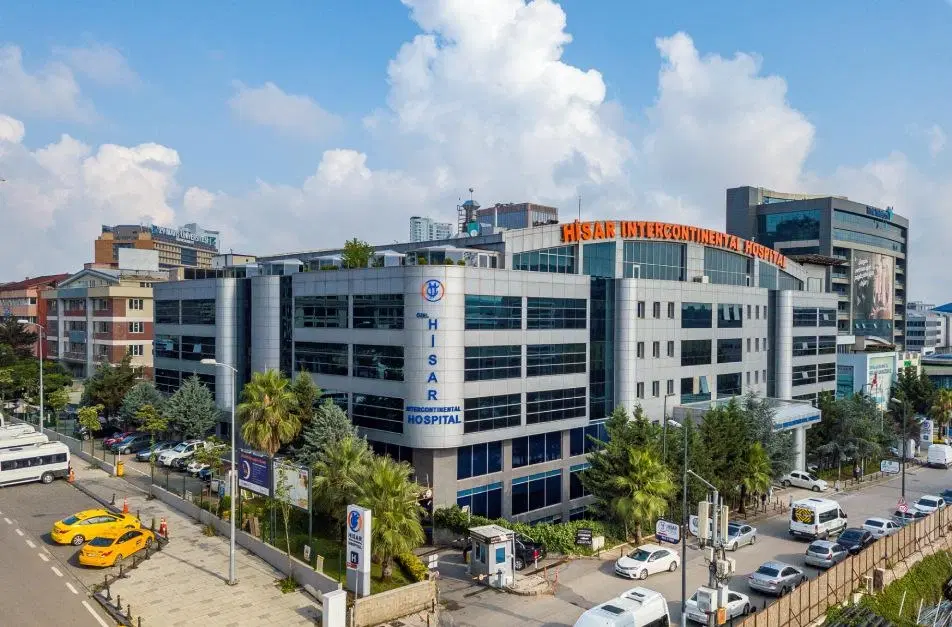

Hisar Intercontinental Hospital
Saray Mah. Siteyolu Cad. No:7, Umraniye, 34768, Istanbul, Turkey

Medical Park Group, Istanbul
Fahrettin Kerim Gokay Cad. Tıbbiye Cd., Kadikoy, Istanbul, Turkey
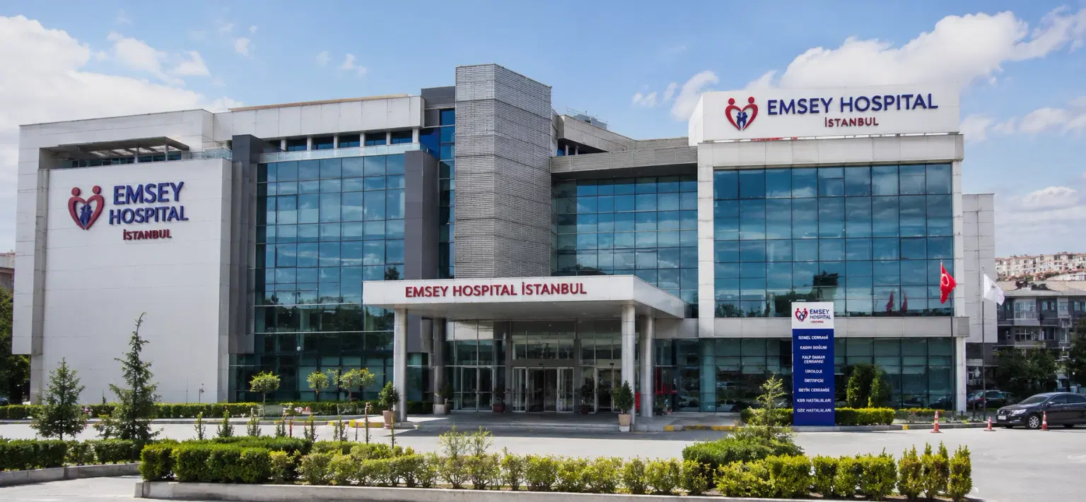
Emsey Hospital, Pendik, Istanbul
Çamlık, Selçuklu Cd. No:22, 34912 Pendik/İstanbul, Türkiye

LIV Hospital, Istanbul
How DivinHeal Helps
We simplify your medical journey by providing comprehensive support and access to world-class healthcare.
Expert Specialist Matching
Connecting you with the world's top-rated medical experts.
Accredited Hospital Network
Access to JCI & NABH certified healthcare facilities.
Complete Travel Coordination
Hassle-free visa, stay, and local transport assistance.
24/7 Personal Care
Dedicated patient advisors supporting you at every step.
Journey Guidance
Full guidance from start to end of the patient treatment journey.
Expert Specialist Matching
Connecting you with the world's top-rated medical experts.
Everything you
need to know today
Browse through these common inquiries to better understand our patient-focused medical platform.
Yes, we work with a variety of insurance providers. Contact our team to verify your coverage.
Yes, we provide secure online consultations with experienced specialists.
Our care coordinators help match you with the most suitable specialist.
Absolutely. Your medical information is protected according to healthcare privacy standards.
Look at six things: accreditation (JCI or NABH), specialty depth, doctor credentials and experience, procedure-specific success rates, international patient support, and technology. DivinHeal's AI-driven matching evaluates every hospital in our accredited partner network on these dimensions and shortlists the best-fit options for your condition, budget, and country.
JCI (Joint Commission International) is the US-based global gold standard for hospital quality, recognised worldwide. NABH is India's national accreditation — accredited by ISQua, the same body that accredits JCI. Both signal independently verified safety and quality. Most of India's leading hospitals hold both.
Yes. All three welcome international patients through structured medical visa programs. India is the most established, treating patients from Africa, the Middle East, and South Asia at 60–80% lower cost. Thailand leads in cosmetic and dental care. The UAE is emerging in oncology and reproductive medicine.
Most patients save 50–80% on treatment costs. Heart bypass costs US $7,000–9,000 in India compared to $70,000–150,000 in the US. IVF costs $3,000–4,500 compared to $12,000–20,000 in the UK. Even after flights, visa, and accommodation, total savings remain 60–70%.
DivinHeal manages your entire non-medical journey: visa invitation letters, medical visa guidance, doctor appointments, teleconsultations, airport pickup, hospital-vetted accommodation for you and your attendant, language interpreters, local transport, cuisine preferences, and post-treatment follow-up — one dedicated coordinator from first enquiry to final follow-up.
You need a valid passport (6+ months validity), a medical visa (M-Visa for India — DivinHeal provides the hospital invitation letter), return flight tickets, recent medical reports and a doctor's referral, current prescription list, and proof of financial means. Any accompanying attendant needs their own passport and MX-Visa.
Still have more questions?
Book a call with our friendly team to learn how DivineHeal simplifies your healthcare journey.