



Vascular Embolisation
5K+ International Patients Treated
40+ Source Countries Served
500+ Accredited Partner Hospitals
98% Patient Satisfaction
80% Average Savings vs USA
10K+ Doctors
NABH, JCI Accredited Hospitals
Free Treatment Plan
Free Consultation with Doctor
5+ Destinations Covered
About Vascular Embolisation
Sources and Guidelines Referenced
This clinical guide is grounded in international interventional radiology guidelines and clinical trials, including: Society of Interventional Radiology (SIR) Quality Improvement Guidelines (2020/2023); Cardiovascular and Interventional Radiological Society of Europe (CIRSE) Standards of Practice (2021); National Institute for Health and Care Excellence (NICE) Interventional Procedures Guidance IPG367 (Uterine Artery Embolisation) and IPG662 (Prostate Artery Embolisation); American Heart Association / American Stroke Association (AHA/ASA) Guidelines for Brain Arteriovenous Malformations (2017); European Association for the Study of the Liver (EASL) Clinical Practice Guidelines on Hepatocellular Carcinoma (2018); and the Spies et al. Fibroid Registry Data Studies (2018).
Vascular Embolisation: A Comprehensive Patient Guide
1. Definition and Medical Identity
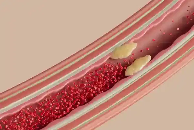
Vascular embolisation, also known as endovascular embolotherapy, is a minimally invasive catheter-based procedure designed to deliberately block or significantly reduce blood flow within specific target blood vessels. Guided by real-time X-ray imaging, interventional radiologists deploy specialized materials to stop internal bleeding, shrink hypervascular tumours, or eliminate abnormal vascular malformations.
The term embolisation derives from the Greek word embolos, meaning a plug or stopper. In modern medical practice, embolotherapy represents a cornerstone of minimally invasive interventional radiology. The primary clinical objective is to achieve selective vessel occlusion (closing off a specific blood vessel) without compromising the blood supply to surrounding healthy tissues. This treatment can be executed as a definitive cure, a preoperative measure to reduce surgical blood loss, or a palliative intervention to control severe symptoms in advanced disease.
Unlike open surgical procedures that require large skin incisions to expose and tie off blood vessels, embolisation works entirely from within the vascular system (an endovascular approach). Using a tiny entry point—typically in the groin or wrist—a specialist navigates microcatheters through the arterial network under real-time image guidance. This technical design allows clinicians to access deep, delicate, or surgically inaccessible regions throughout the human body with exceptional precision.
2. The Underlying Condition or Need
Vascular embolisation addresses vascular pathologies characterized by uncontrolled bleeding, abnormal vessel connections, or excessive tissue proliferation funded by blood vessels. Unmanaged arterial bleeding or hypervascular growth can cause life-threatening haemorrhage, organ dysfunction, or progressive tissue destruction, requiring targeted interruption of regional vascular perfusion.
Under normal physiological conditions, the human circulatory system maintains precise control over blood flow, delivering oxygen and nutrients while containing intravascular pressure. However, structural defects, traumatic injuries, or pathologically altered tissues can disrupt this balance:
- Acute Arterial Haemorrhage: Traumatic injuries, pelvic fractures, gastrointestinal ulcers, or surgical complications can rupture arterial walls, producing rapid internal blood loss. Without prompt control, acute bleeding can lead to hypovolaemic shock (dangerously low blood pressure from blood loss) and organ failure.
- Hypervascular Benign and Malignant Tumours: Conditions such as symptomatic uterine fibroids, benign prostatic hyperplasia, or primary liver cancer (hepatocellular carcinoma) develop dense, abnormal networks of blood vessels. These vessels supply rapid cellular growth and, in the case of fibroids or prostate enlargement, produce painful structural compression on surrounding organs.
- Vascular Malformations and Aneurysms: Congenital or acquired vascular lesions—including arteriovenous malformations (AVMs) and visceral or intracranial aneurysms—feature fragile, high-pressure vessel connections that carry a permanent risk of spontaneous rupture and severe bleeding.
When left untreated, these conditions can cause severe outcomes ranging from chronic debilitating pain and severe anaemia to catastrophic, sudden internal collapse. Vascular embolisation provides a rapid, targeted therapeutic solution that directly halts pathologic blood flow at its biological source.
3. How the Treatment Works — Mechanism
Vascular embolisation works by introducing mechanical barriers or biological sclerosing agents directly into target arterial or venous channels. Once deployed, these agents induce targeted mechanical occlusion, local blood stasis, thrombosis, and secondary fibrous tissue formation, effectively cutting off oxygen and nutrient delivery while preserving surrounding healthy tissues.
The physiological cascade of embolotherapy follows a defined biological process:
First, the interventional radiologist navigates a microcatheter (a flexible catheter measuring less than one millimetre in outer diameter) into the specific branch vessel supplying the lesion. Fluoroscopy—a continuous real-time X-ray technique—combined with contrast medium injection allows exact visualization of blood vessel architecture through digital subtraction angiography (DSA).
Once correct catheter placement is verified, the chosen embolic material is delivered. The primary biological mechanism depends on the embolic agent selected:
- Mechanical Occlusion: Metallic coils or vascular plugs physically block macrovascular blood flow. The metal structure acts as a physical barrier that slows blood velocity, triggering the body's natural coagulation cascade.
- Microvascular Stasis: Microparticles (such as tris-acryl gelatin microspheres or polyvinyl alcohol) travel distally into small arteriole beds, physically wedging into tiny vessels. This produces immediate localized blood stasis and ischemic tissue necrosis.
- Chemical Sclerosis and Polymerization: Liquid embolic agents (such as n-butyl cyanoacrylate glue or ethylene vinyl alcohol copolymer) contact blood or physiological saline and rapidly solidify or cause immediate vessel wall endothelial damage, creating an instantaneous permanent cast of the vascular bed.
Following primary occlusion, local blood stagnation triggers natural intravascular thrombosis (blood clotting). Over subsequent days to weeks, inflammatory cells enter the trapped clot, replacing it with permanent fibrous scar tissue. In hypervascular tumours or fibroids, this sudden loss of blood supply leads to ischemia, cell death, and gradual tissue shrinkage over several months, while normal surrounding tissues remain protected by collateral blood vessels.
4. Types and Variations
Vascular embolisation encompasses multiple sub-types based on the clinical objective, targeted organ, and type of embolic agent utilized. Variations range from temporary occlusions using gelfoam to permanent occlusions using metallic coils, microparticles, liquid embolic agents, or chemoembolisation mixtures tailored to specific vessel anatomical structures.
Embolic agents are broadly classified by their duration of action (temporary versus permanent) and physical state (solid, particulate, or liquid):
| Embolic Category | Primary Materials | Mechanism & Duration | Primary Clinical Indications |
|---|---|---|---|
| Temporary Particulate Agents | Gelatin sponge (Gelfoam) | Resorbable mechanical occlusion; degrades within 2 to 6 weeks | Traumatic visceral bleeding, temporary control prior to surgery, postpartum haemorrhage |
| Permanent Microparticles | Polyvinyl alcohol (PVA), Tris-acryl gelatin microspheres | Permanent distal arteriolar occlusion; mechanical blockade and thrombosis | Uterine Artery Embolisation (UAE), Prostate Artery Embolisation (PAE), hypervascular tumours |
| Mechanical Devices | Platinum/stainless steel coils, Nitinol vascular plugs | Permanent focal macrovascular occlusion; physically blocks vessel lumen | Visceral aneurysms, arteriovenous fistulas, direct arterial trunk occlusion |
| Liquid Embolic Agents | Ethylene vinyl alcohol copolymer (Onyx/SQUID), N-butyl cyanoacrylate (NBCA glue) | Permanent liquid casting; polymerizes or precipitates upon blood contact | High-flow arteriovenous malformations (AVMs), hypervascular cranial/spinal lesions |
| Chemoembolisation (TACE) | Drug-eluting beads (DEB-TACE) loaded with doxorubicin or cisplatin | Combined targeted chemotherapy delivery with mechanical ischemia | Hepatocellular carcinoma (primary liver cancer), liver metastases |
| Radioembolisation (TARE/SIRT) | Yttrium-90 (Y90) resin or glass microspheres | Internal radiation delivery combined with microvascular embolisation | Unresectable liver tumors, advanced hepatic malignancies |
The choice of variation depends on whether the vessel occlusion needs to be permanent or temporary, the size and velocity of blood flow within the vessel, and whether localized delivery of chemotherapy or radiation is required alongside vascular blockage.
5. Who the Treatment Is For — Indications
Vascular embolisation is indicated for patients with acute or chronic arterial haemorrhage, hypervascular benign or malignant tumours, vascular malformations, visceral aneurysms, uterine fibroids, or benign prostatic hyperplasia. Candidates undergo pre-procedural imaging and clinical risk stratification to confirm vascular anatomical suitability for endovascular intervention.
Key clinical indications supported by major clinical guidelines include:
- Uterine Fibroids (Uterine Artery Embolisation - UAE): Recommended by NICE (IPG367) and ACOG guidelines as a safe, effective organ-preserving alternative to hysterectomy or myomectomy for women with heavy menstrual bleeding and pelvic bulk symptoms.
- Benign Prostatic Hyperplasia (Prostate Artery Embolisation - PAE): Supported by NICE (IPG662) and SIR guidelines for men with moderate-to-severe lower urinary tract symptoms who are poor surgical candidates or wish to preserve sexual function.
- Acute Traumatic and Postoperative Haemorrhage: Indicated by CIRSE standards as the primary first-line treatment for unstable pelvic fractures, active retroperitoneal bleeding, and blunt abdominal organ trauma (splenic, hepatic, or renal lacerations).
- Gastrointestinal Bleeding: Selected for upper or lower GI bleeding refractory to endoscopic interventions, rapidly arresting arterial blood loss without open abdominal surgery.
- Malignant Liver Tumours (TACE/TARE): Recommended by the European Association for the Study of the Liver (EASL) as standard-of-care for intermediate-stage hepatocellular carcinoma (Child-Pugh Class A/B).
- Vascular Lesions and Aneurysms: Utilized under AHA/ASA guidelines to eliminate brain and visceral arteriovenous malformations, pelvic varicoceles, and renal or splenic artery aneurysms.
Diagnostic workup requires cross-sectional imaging (CT Angiography or MR Angiography) to map arterial supply, confirm catheter accessibility, and exclude anatomical anomalies before scheduling the procedure.
6. Who the Treatment Is NOT For — Contraindications
Vascular embolisation is contraindicated in patients with severe uncorrectable coagulopathies, active unmanaged systemic infections, severe renal insufficiency precluding iodinated contrast use, or vascular anatomy that prevents safe catheter navigation without risking critical non-target ischemia to essential tissue structures.
Contraindications are divided into absolute and relative clinical factors:
Absolute Contraindications
- Uncontrollable Systemic Sepsis or Active Local Infection: Embolising tissue in the presence of active bacteremia significantly increases the risk of infected tissue necrosis and abscess formation.
- Irreversible Severe Anaphylactic Reaction to Contrast Media: When alternative non-ionic contrast agents or carbon dioxide ($CO_2$) angiography cannot be safely substituted.
- Anatomical Inaccessibility: Severe arterial tortuosity or complete vessel occlusion that physically prevents microcatheter navigation to the target site.
- Pregnancy (for elective non-emergent procedures): Due to radiation exposure and potential alteration of uterine blood flow. Emergency life-saving maternal embolisation remains an exception.
Relative Contraindications
- Severe Renal Dysfunction (eGFR < 30 mL/min/1.73m²): Requires volume hydration, minimal contrast dosing, or alternative contrast agents ($CO_2$) to prevent contrast-induced acute kidney injury.
- Severe Uncorrected Coagulopathy or Thrombocytopenia: Platelet counts under 50,000/µL or INR greater than 1.5–1.8 increase vascular puncture risks and require pre-procedure blood product transfusion.
- Borderline Organ Failure: In liver chemoembolisation, advanced hepatic cirrhosis (Child-Pugh Class C) significantly increases the risk of acute post-procedural liver failure.
7. Alternatives and Clinical Comparison
Alternatives to vascular embolisation include open or laparoscopic surgical vessel ligation, surgical tissue resection, conservative medical therapies, endoscopic interventions, and stereotactic radiosurgery. The selection depends on patient surgical candidacy, vascular accessibility, underlying pathology, organ preservation priorities, and acute haemodynamic stability.
The following table compares embolisation against established surgical and medical alternatives:
| Treatment Approach | Invasiveness & Access | Primary Advantages | Clinical Trade-offs & Limitations |
|---|---|---|---|
| Vascular Embolisation | Minimally invasive; 2mm skin puncture (femoral/radial) | No general anaesthesia required; rapid recovery; organ-preserving; low blood loss | Post-embolisation syndrome; potential risk of non-target embolisation; relies on fluoroscopy |
| Open Surgical Resection / Ligation | Invasive; full surgical incision under general anaesthesia | Direct anatomical visualization; definitive immediate removal of entire lesion | Higher blood loss; longer hospital stay; increased infection risk; significant scar tissue |
| Laparoscopic / Robotic Surgery | Moderately invasive; multiple small abdominal ports | Faster recovery than open surgery; excellent direct organ visualization | Requires general anaesthesia; abdominal insufflation; limited access in severe adhesions |
| Pharmacotherapy (e.g., Hormonal/Antifibrinolytic) | Non-invasive; oral or injectable medications | No surgical or procedural risks; widely accessible | Symptoms often recur upon stopping therapy; side effects with long-term medication use |
| Stereotactic Radiosurgery (for AVMs) | Non-invasive focused radiation delivery | No mechanical puncture; targets deep non-operable intracranial lesions | Delayed onset of action (1–3 years for complete occlusion); radiation exposure |
Interdisciplinary tumor boards and clinical panels evaluate each patient's specific anatomical features, health status, and treatment goals to select the optimal treatment method.
8. Pre-Treatment Phase
The pre-treatment phase includes diagnostic vascular imaging, comprehensive lab evaluations, and clinical risk assessments. Patients undergo contrast renal clearance testing, medication adjustments for anticoagulants, pre-procedure hydration, and formal informed consent discussions regarding potential benefits, technical risks, and post-procedure recovery expectations.
Once a patient is referred for embolisation, a structured evaluation process begins:
1. Initial Clinical Consultation and History
An interventional radiologist evaluates the patient's medical background, focusing on cardiovascular health, allergy history (especially to iodinated contrast media or shellfish), prior surgical history, and current medications.
2. Diagnostic Imaging and Mapping
High-resolution cross-sectional imaging—such as multiphase CT Angiography (CTA) or Magnetic Resonance Angiography (MRA)—is reviewed. This creates a detailed three-dimensional map of the patient's arterial branching, identifying targeted vessels and warning of potential anatomical variations.
3. Laboratory Diagnostic Workup
- Renal Function Panel: Measuring serum creatinine and calculating estimated glomerular filtration rate (eGFR) to ensure safe clearance of iodinated contrast agents.
- Coagulation Screening: Checking International Normalised Ratio (INR), Prothrombin Time (PT), Activated Partial Thromboplastin Time (aPTT), and platelet counts to minimize puncture site bleeding risks.
- Complete Blood Count (CBC): Baseline hemoglobin and hematocrit to evaluate prior blood loss.
4. Medication Management and Fasting
Anticoagulants (such as warfarin, rivaroxaban, or apixaban) and antiplatelet drugs (such as clopidogrel) are temporarily paused according to SIR consensus protocols. Patients are instructed to fast for 6 to 8 hours prior to the procedure. Intravenous fluid hydration is started 2 to 4 hours before the procedure in patients with borderline renal function.
9. The Procedure — Step-by-Step Clinical Detail
The procedure involves sterile vascular access, fluoroscopic catheter navigation, microcatheter super-selection of target vessels, selective contrast angiography, embolic agent deployment, post-embolic angiographic verification, and vascular access site closure. Performed under local anaesthesia with conscious sedation, the intervention typically lasts between 60 and 180 minutes.
Step 1: Patient Preparation and Sedation
The patient is positioned on an interventional radiology fluoroscopy table. Standard monitoring equipment (ECG, pulse oximetry, non-invasive blood pressure) is attached. Local anaesthesia (typically 1% or 2% lidocaine) is injected into the selected access site—either the common femoral artery in the groin or the radial artery in the wrist. Intravenous conscious sedation (commonly midazolam and fentanyl) is administered to ensure comfort while keeping the patient awake enough to follow breathing instructions.
Step 2: Vascular Access (Seldinger Technique)
Using ultrasound guidance, the interventional radiologist punctures the target artery with a small needle. A fine guide wire is passed through the needle into the arterial lumen, over which a flexible vascular sheath (5 to 6 French size, approximately 1.6 to 2.0 mm diameter) is placed. This sheath acts as a protected entry portal throughout the procedure.
Step 3: Diagnostic Angiography and Catheter Navigation
A primary diagnostic catheter is advanced over a guidewire through the aorta and positioned at the main arterial trunk supplying the target organ (such as the internal iliac artery for fibroids or the celiac/hepatic artery for liver tumours). Iodinated contrast medium is injected through the catheter while continuous fluoroscopic X-rays are captured, producing clear digital subtraction angiography (DSA) images of the blood vessels.
Step 4: Microcatheter Super-Selection
Through the primary catheter, a coaxial microcatheter (1.7 to 2.7 French size) and microguidewire are carefully navigated into the microvascular branches directly feeding the pathological lesion. This precise positioning—termed super-selective catheterization—ensures embolic materials are delivered only to diseased tissue while protecting healthy adjacent vessel branches.
Step 5: Embolic Agent Deployment
Under continuous fluoroscopy monitoring, the chosen embolic agent (microparticles, liquid polymer, or metallic coils) is infused through the microcatheter. The radiologist monitors contrast movement in real-time. Embolic delivery continues until target vessel blood flow slows to a complete stop (end-point stasis).
Step 6: Completion Angiography and Hemostasis
A final diagnostic angiogram is performed from the main catheter to confirm complete obliteration of the target vessel and verify that normal blood flow to non-target tissue remains intact. The catheters and sheath are removed. Hemostasis at the arterial access site is achieved using an internal vascular closure device (such as a collagen plug or suture-based system) or manual compression applied for 15 to 20 minutes.
10. Immediate Post-Procedure Period
The immediate post-procedure period focuses on arterial access site monitoring, bed rest, haemodynamic observation, renal hydration, and pain control. Patients remain in a specialized recovery unit for four to twenty-four hours to detect potential early access-site bleeding, distal pulse changes, or acute post-embolisation pain symptoms.
Key components of immediate post-procedure care include:
- Bed Rest and Access Site Positioning: If femoral artery access was used, the patient must lie flat with the affected leg straight for 2 to 6 hours to allow the puncture site to seal reliably. Radial wrist access allows immediate sitting and ambulation with a compression wrist band.
- Vascular and Neurological Checks: Nursing staff perform frequent monitoring of peripheral pulses (dorsalis pedis or radial pulse), puncture site check for swelling or hematoma formation, and blood pressure monitoring every 15 minutes for the first hour.
- Pain and Symptom Management: As embolic tissue ischemia develops, patients may experience localized pain. Intravenous patient-controlled analgesia (PCA) or oral nonsteroidal anti-inflammatory drugs (NSAIDs) and opioids are administered according to pain management protocols.
- Intravenous Hydration: IV fluids are continued to enhance renal clearance of contrast medium and reduce the risk of contrast-induced nephropathy.
- Discharge Assessment: Patients receiving elective procedures (such as UAE or PAE) are frequently discharged the same day or following an overnight stay once stable ambulation, adequate oral pain control, and normal voiding are demonstrated.
11. Recovery — Short and Long Term
Recovery spans initial physical rest over two to seven days, followed by gradual tissue remodelling and symptom resolution over four to twelve weeks. Mild post-embolisation symptoms subside within days, while complete vessel obliteration and organ or tissue shrinkage are monitored through scheduled follow-up cross-sectional imaging.
Short-Term Recovery (Days 1 to 14)
During the first week, patients frequently experience a cluster of inflammatory symptoms called post-embolisation syndrome (PES). PES is an expected response to tissue ischemia and cell breakdown, not an infection. Symptoms include:
- Low-grade fever (typically under 38.5°C / 101.3°F)
- Localized pelvic, abdominal, or back pain
- General fatigue, malaise, and mild nausea
PES typically peaks between 24 and 48 hours post-procedure and resolves within 5 to 7 days. Management relies on scheduled oral NSAIDs, short-term antiemetics, and adequate fluid intake. Most patients return to light desk work and normal daily activities within 7 to 10 days.
Long-Term Recovery and Follow-up Timeline (Weeks 2 to 52)
- Week 2 to 4: Subacute inflammatory symptoms disappear. Patients can resume routine exercise and regular physical activity. Access site puncture marks heal completely.
- Month 1 to 3: Initial follow-up appointment with the interventional radiologist. Contrast-enhanced MRI or ultrasound is routinely performed (e.g., in UAE or PAE) to measure volume reduction in targeted tissues and verify complete vascular occlusion.
- Month 6 to 12: Maximal clinical benefit and tissue remodeling are achieved. For fibroids and prostate enlargement, volume reductions between 40% and 70% are typically documented by 6 months, accompanied by significant symptom improvement.
12. Risks, Side Effects, and Complications
While vascular embolisation is generally safe and minimally invasive, potential complications range from mild post-embolisation syndrome and access site hematomas to rare severe events like non-target embolisation, vascular dissection, contrast-induced acute kidney injury, or tissue necrosis requiring secondary intervention.
The overall complication rate across major interventional radiology registries (such as the SIR Quality Improvement Guidelines) is low, with major complications occurring in less than 2% to 3% of elective cases.
| Severity Level | Complication / Side Effect | Estimated Frequency | Clinical Presentation & Management |
|---|---|---|---|
| Common / Mild | Post-Embolisation Syndrome (PES) | 40% – 80% | Low-grade fever, pelvic/abdominal pain, nausea, malaise. Self-limiting; managed with oral analgesics and antiemetics for 3–7 days. |
| Common / Mild | Access site ecchymosis (bruising) | 10% – 20% | Localized discoloration at groin or wrist puncture. Resolves spontaneously over 1–2 weeks without intervention. |
| Uncommon / Moderate | Access site haematoma / Pseudoaneurysm | 1% – 3% | Painful swelling at puncture site. Small haematomas resolve naturally; pseudoaneurysms managed with compression or ultrasound-guided thrombin injection. |
| Uncommon / Moderate | Contrast-Induced Acute Kidney Injury | 1% – 2% | Transient rise in serum creatinine due to contrast clearance stress. Prevented and managed with IV hydration; higher risk in pre-existing renal disease. |
| Rare / Serious | Non-Target Embolisation | < 1% | Accidental embolization of healthy adjacent arteries leading to skin, bowel, or nerve ischemia. Minimised by microcatheter techniques and fluoroscopic monitoring. |
| Rare / Serious | Arterial Dissection or Perforation | < 0.5% | Catheter-induced tear in vessel wall. Treated intra-procedurally via balloon angioplasty, covered stent placement, or focal micro-coiling. |
| Rare / Serious | Infection / Tissue Abscess Formation | < 0.5% | Infection in ischemic devascularized tissue. Presents with late high fever and severe pain; treated with systemic antibiotics or percutaneous drainage. |
Warning signs requiring urgent medical evaluation after discharge include fever exceeding 38.5°C (101.3°F), rapidly expanding swelling or severe pain at the groin or wrist access site, cold or pale extremities, or sudden shortness of breath.
13. Lifestyle and Behavioural Considerations
Post-treatment lifestyle recommendations focus on access site protection, temporary physical exertion restrictions, adequate hydration to facilitate contrast renal clearance, and adherence to prescribed post-procedure medication protocols including pain management and occasional short-term antiplatelet therapy where clinically indicated.
Pre-Procedure Optimization
- Smoking Cessation: Halting tobacco use at least 2 weeks prior improves peripheral vascular health, enhances tissue healing, and decreases cardiopulmonary risks during sedation.
- Hydration: Increasing oral fluid intake 24 to 48 hours prior to intervention helps optimize circulating intravascular volume and supports post-contrast kidney function.
Post-Procedure Activity Guidelines
- Lifting and Straining: Patients must avoid heavy lifting (greater than 5 kilograms / 10 pounds) and strenuous physical exertion for 5 to 7 days following femoral access to prevent delayed puncture site bleeding or pseudoaneurysm formation.
- Bathing and Hygiene: Puncture sites should be kept clean and dry. Showering is permitted after 24 to 48 hours, but soaking in baths, hot tubs, or swimming pools should be avoided for 7 to 10 days until the skin entry site is completely closed.
- Dietary Adjustments: High-fiber foods and adequate fluid intake are recommended during the first week to prevent constipation, which can worsen pelvic pain or strain arterial access recovery sites.
14. How Outcomes Are Measured
Clinical outcomes are evaluated by technical success (complete targeted vessel occlusion), clinical success (symptom relief or cessation of bleeding), and radiological follow-up using contrast-enhanced ultrasound, CT angiography, or MRI to verify sustained vascular blockage and tissue volume reduction.
The definition of successful outcome varies by underlying indication:
- Technical Success: Achieved when the interventional radiologist successfully accesses the target vessel and delivers embolic material to achieve complete stasis of target blood flow, as verified by post-deployment contrast angiography. International registry data (SIR 2023) show technical success rates consistently exceeding 95% across major embolisation indications.
- Clinical Success: Defined as meaningful reduction or elimination of symptoms. In emergency haemorrhage, clinical success means rapid stabilization of blood pressure without requiring surgical ligation. In uterine artery embolisation, clinical success is measured using validated symptom severity questionnaires (such as the UFS-QOL score), showing durable clinical relief in 85% to 90% of patients at 5-year follow-up (Spies et al.).
- Radiological Success: Evaluated on follow-up cross-sectional imaging (MRI or CT) by measuring the percentage reduction in target lesion volume (e.g., fibroid or prostate volume reduction) and demonstrating complete absence of contrast enhancement inside the treated lesion (indicating sustained lack of blood flow).
If incomplete embolisation occurs or collateral blood supply develops over time, a secondary repeat embolisation or transition to surgical management can be safely evaluated by the care team.
15. Recent Advances and Current Standard of Care
Modern endovascular embolotherapy leverages advanced cone-beam computed tomography (CBCT) navigation, microcatheter technology, drug-eluting beads, and bioresorbable embolic materials. Current guidelines emphasize multidisciplinary team decision-making, super-selective technique application, and organ-sparing vascular approaches across both elective and emergency clinical presentations.
Over the past decade, several technical innovations have reshaped standard interventional radiology practice:
- Cone-Beam CT (CBCT) and Vessel Tracking Software: Interventional suites now utilize C-arm CT technology that generates three-dimensional angiographic images in the procedure room. Automated vessel-tracking algorithms guide microcatheters into tiny feeder vessels, reducing procedure times and radiation exposure.
- Calibrated Bioresorbable and Hydrogel Microspheres: Advanced microspheres provide uniform arterial occlusion and can absorb specific chemo-therapeutic agents or slowly dissolve over pre-programmed timeframes, reducing permanent foreign body exposure.
- Radial Artery Access Approach: Adapting techniques from interventional cardiology, interventional radiologists increasingly use the transradial access route (wrist access) for abdominal and pelvic embolisations. Clinical trials show transradial access reduces access-site bleeding complications, improves patient comfort, and allows immediate post-procedure walking.
- AI-Assisted Treatment Planning: Emerging artificial intelligence applications assist in mapping complex arterial supply to hypervascular tumours, optimizing embolic dosing and predicting non-target occlusion risks before microcatheter insertion.
16. Common Myths and Misconceptions
Unfounded myths surrounding vascular embolisation often confuse catheter-based procedures with open surgery or assume that blocking blood flow permanently damages entire organs. Evidence-based facts demonstrate precise super-selective targeting that protects healthy surrounding tissue while achieving therapeutic endovascular goals.
Myth: Embolisation cuts off blood flow to the entire organ, causing organ necrosis.
Reality: Microcatheter technology allows highly selective targeting of only the pathologically altered blood vessels supplying diseased tissue. Surrounding healthy organ structures continue to receive adequate blood supply through normal collateral vessel networks (CIRSE Standards of Practice 2021).
Myth: Embolisation is an experimental treatment option.
Reality: Embolotherapy has been practiced for over four decades. It is established in international guidelines, including those from NICE, SIR, ACOG, and EASL, as a standard-of-care primary therapy for conditions ranging from traumatic bleeding to uterine fibroids.
Myth: Embolisation requires major general anaesthesia and prolonged hospital stays.
Reality: Most embolisation procedures are performed under local anaesthesia with conscious sedation. Many patients are discharged the same day or after a short 24-hour observation period.
Myth: Embolic materials can travel freely throughout the body forever.
Reality: Embolic agents are sized specifically to match target vessel diameters. Once wedged into target arterioles or delivered into selected vessel segments, they remain locked in place within the localized clot and fibrous tissue matrix.
Myth: Uterine artery embolisation causes immediate menopause in all women.
Reality: According to ACOG clinical guidelines, the overall risk of permanent ovarian failure following UAE is under 2% in women under age 40, though the risk increases in women over age 45 as natural menopause approaches.
Myth: If embolisation fails, open surgery is no longer possible.
Reality: Embolisation does not alter non-vascular anatomical boundaries. If a condition requires further management, conventional laparoscopic or open surgical procedures can still be safely performed.
17. Frequently Asked Questions
What is vascular embolisation?
Vascular embolisation is a minimally invasive catheter-based treatment performed by interventional radiologists. Under real-time imaging, specialists navigate microcatheters into specific blood vessels to deliver occlusive materials (coils, particles, or liquids), stopping internal bleeding or starving abnormal tissues and tumours of their blood supply.
Is vascular embolisation considered major surgery?
No. Vascular embolisation is a minimally invasive endovascular procedure, not open surgery. It requires no large skin incisions, relies on a tiny 2 mm entry point in the groin or wrist, and is typically performed under local anaesthesia with light sedation.
How long does the embolisation procedure take?
Most vascular embolisation procedures take between 60 and 180 minutes, depending on the complexity of the vascular anatomy, the number of target vessels being treated, and the specific embolic agents deployed.
Will I feel pain during vascular embolisation?
Patients do not feel catheters moving within the vascular system because blood vessel walls lack pain receptors. Local anaesthesia numbs the skin access site, and IV sedation keeps patients relaxed. Some localized cramping or pain may develop shortly after embolic delivery, which is managed with intravenous analgesics.
What is post-embolisation syndrome?
Post-embolisation syndrome (PES) is a self-limiting inflammatory response triggered by tissue ischemia following vessel blockage. Symptoms include low-grade fever, pelvic or abdominal discomfort, nausea, and fatigue. PES typically peaks within 48 hours and resolves in 3 to 7 days with oral medications.
How long do I need to stay in the hospital?
Hospital stays range from 4 to 24 hours. Many elective embolisation procedures (such as uterine artery embolisation or prostate artery embolisation) are routinely managed as outpatient or single-overnight-stay procedures.
Are embolic agents permanent?
Embolic agents can be permanent or temporary, depending on the medical need. Metallic coils, microspheres, and liquid polymers provide permanent vessel occlusion, whereas gelatin sponge (Gelfoam) provides temporary blockage that naturally dissolves within 2 to 6 weeks.
How quickly will I see results after embolisation?
For acute arterial bleeding, blood loss halts immediately during the procedure. For structural lesions such as uterine fibroids or prostate enlargement, symptom relief begins within 2 to 4 weeks, with progressive tissue shrinkage continuing over 3 to 6 months.
When can I return to work and normal activity?
Most patients resume light desk work and daily routine activities within 3 to 7 days post-procedure. Strenuous physical exercise and heavy lifting should be paused for 7 to 10 days to allow complete healing of the arterial access site.
Can embolic coils or particles move to other parts of my body?
Inadvertent movement, known as non-target embolisation, is rare (occurring in less than 1% of procedures). Interventional radiologists use continuous fluoroscopy and precise sizing calibrations to ensure agents lodge securely within target vessels.
Is radiation exposure from fluoroscopy safe?
Interventional radiologists follow strict principles to minimize radiation doses during fluoroscopy. Modern equipment utilizes pulsed imaging and digital subtraction software to achieve diagnostic visualization with minimal exposure.
How do I know if the embolisation worked?
Treatment success is confirmed through clinical symptom improvement and follow-up cross-sectional imaging (contrast-enhanced MRI, CT, or ultrasound) scheduled at 1 to 3 months post-procedure to verify tissue volume reduction and complete loss of blood flow in target vessels.
Booking With DIVINHEAL
Get a free consultation to understand your treatment options
Cost Calculator
I know my treatment - show me cost from 3 hospitals
Plan My Journey
Tell us your condition and budget - our AI matches the right destination, hospital and doctor and visa pathway

Booking With DIVINHEAL
Get a free consultation to understand your treatment options
Cost Calculator
I know my treatment - show me cost from 3 hospitals
Plan My Journey
Tell us your condition and budget - our AI matches the right destination, hospital and doctor and visa pathway
Meet Our Medical Specialists
Hospitals
NABH & JCI Accredited Hospitals in India,Turkey, Thailand & UAE.
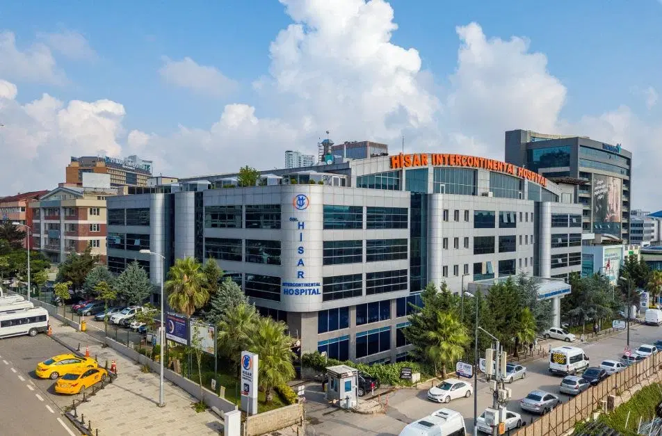

Hisar Intercontinental Hospital
Saray Mah. Siteyolu Cad. No:7, Umraniye, 34768, Istanbul, Turkey

Medical Park Group, Istanbul
Fahrettin Kerim Gokay Cad. Tıbbiye Cd., Kadikoy, Istanbul, Turkey
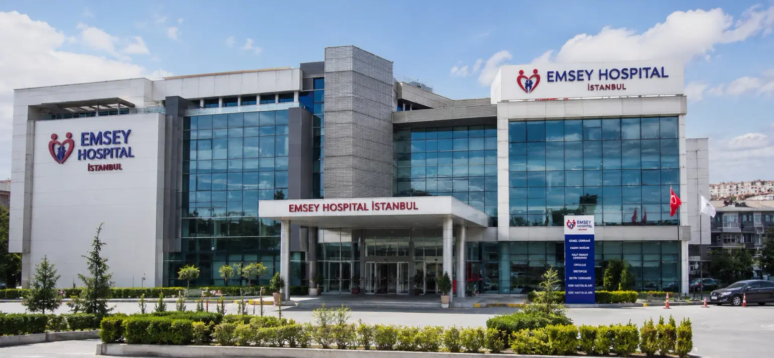
Emsey Hospital, Pendik, Istanbul
Çamlık, Selçuklu Cd. No:22, 34912 Pendik/İstanbul, Türkiye
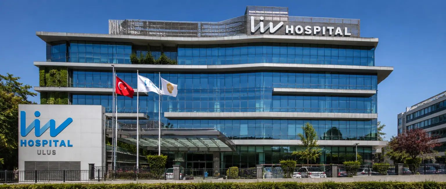
LIV Hospital, Istanbul
How DivinHeal Helps
We simplify your medical journey by providing comprehensive support and access to world-class healthcare.
Expert Specialist Matching
Connecting you with the world's top-rated medical experts.
Accredited Hospital Network
Access to JCI & NABH certified healthcare facilities.
Complete Travel Coordination
Hassle-free visa, stay, and local transport assistance.
24/7 Personal Care
Dedicated patient advisors supporting you at every step.
Journey Guidance
Full guidance from start to end of the patient treatment journey.
Expert Specialist Matching
Connecting you with the world's top-rated medical experts.
Everything you
need to know today
Browse through these common inquiries to better understand our patient-focused medical platform.
Yes, we work with a variety of insurance providers. Contact our team to verify your coverage.
Yes, we provide secure online consultations with experienced specialists.
Our care coordinators help match you with the most suitable specialist.
Absolutely. Your medical information is protected according to healthcare privacy standards.
Look at six things: accreditation (JCI or NABH), specialty depth, doctor credentials and experience, procedure-specific success rates, international patient support, and technology. DivinHeal's AI-driven matching evaluates every hospital in our accredited partner network on these dimensions and shortlists the best-fit options for your condition, budget, and country.
JCI (Joint Commission International) is the US-based global gold standard for hospital quality, recognised worldwide. NABH is India's national accreditation — accredited by ISQua, the same body that accredits JCI. Both signal independently verified safety and quality. Most of India's leading hospitals hold both.
Yes. All three welcome international patients through structured medical visa programs. India is the most established, treating patients from Africa, the Middle East, and South Asia at 60–80% lower cost. Thailand leads in cosmetic and dental care. The UAE is emerging in oncology and reproductive medicine.
Most patients save 50–80% on treatment costs. Heart bypass costs US $7,000–9,000 in India compared to $70,000–150,000 in the US. IVF costs $3,000–4,500 compared to $12,000–20,000 in the UK. Even after flights, visa, and accommodation, total savings remain 60–70%.
DivinHeal manages your entire non-medical journey: visa invitation letters, medical visa guidance, doctor appointments, teleconsultations, airport pickup, hospital-vetted accommodation for you and your attendant, language interpreters, local transport, cuisine preferences, and post-treatment follow-up — one dedicated coordinator from first enquiry to final follow-up.
You need a valid passport (6+ months validity), a medical visa (M-Visa for India — DivinHeal provides the hospital invitation letter), return flight tickets, recent medical reports and a doctor's referral, current prescription list, and proof of financial means. Any accompanying attendant needs their own passport and MX-Visa.
Still have more questions?
Book a call with our friendly team to learn how DivineHeal simplifies your healthcare journey.