



Wisdom Tooth Removal (Impacted)
5K+ International Patients Treated
40+ Source Countries Served
500+ Accredited Partner Hospitals
98% Patient Satisfaction
80% Average Savings vs USA
10K+ Doctors
NABH, JCI Accredited Hospitals
Free Treatment Plan
Free Consultation with Doctor
5+ Destinations Covered
About Wisdom Tooth Removal (Impacted)
Sources and Guidelines Referenced
The clinical recommendations, surgical protocols, and safety data in this guide are derived from published guidelines and peer-reviewed literature from major surgical and health organizations, including: the American Association of Oral and Maxillofacial Surgeons (AAOMS 2021 Clinical Practice Guidelines), the National Institute for Health and Care Excellence (NICE TA1 Guidelines on Wisdom Teeth Extraction), the Royal College of Surgeons of England (RCS 2020 Management of Impacted Third Molars), and landmark studies including Ghaeminia et al. (Cochrane Database of Systematic Reviews, 2020) and Renton et al. (British Dental Journal, 2012).
Wisdom Tooth Removal (Impacted): A Comprehensive Patient Guide
1. Definition and Medical Identity
Wisdom tooth removal for impacted teeth is a specialized surgical procedure to extract third molars that cannot fully erupt into the oral cavity. Known clinically as third molar odontectomy, this procedure belongs to oral and maxillofacial surgery and aims to prevent local tissue infection, structural tooth damage, and jaw disease.
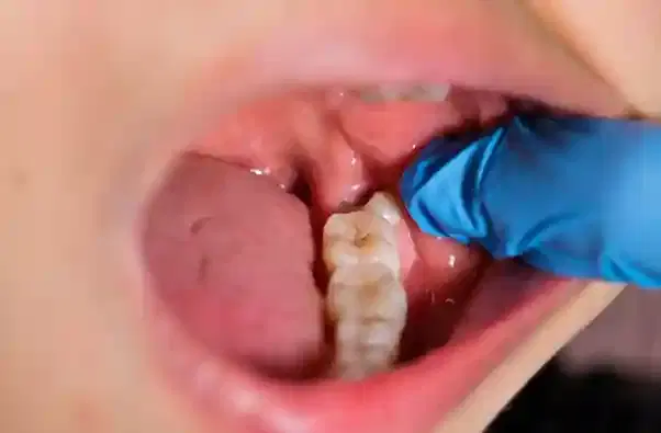
Third molars are the last teeth to form in the human dentition, located at the far posterior ends of the upper maxilla and lower mandible. An impacted tooth is defined as a tooth that is blocked from attaining its correct positional alignment by soft tissue, neighboring teeth, or bone. Impactions are broadly divided into soft tissue impaction, where the tooth crown has cleared the jawbone but remains covered by oral mucosa, and bony impaction, where the tooth remains partially or completely encased within the jawbone.
2. The Underlying Condition or Need
Impacted wisdom teeth develop primarily due to an evolutionary mismatch between human jaw size and tooth number, resulting in inadequate space for proper eruption. Unextracted impacted teeth can trigger localized chronic inflammation, severe infection, structural destruction of nearby second molars, and formation of pathological fluid-filled sacs called cysts.
When a wisdom tooth partially breaches the gum tissue, it creates a small mucosal flap called an operculum. Food debris and oral bacteria easily accumulate underneath this flap, forming an environment that is impossible to clean with routine brushing. This leads to pericoronitis, an acute or chronic soft-tissue infection characterized by severe localized pain, localized facial swelling, foul taste, and trismus (inability to fully open the mouth).
In addition to infection, an angled wisdom tooth pressing against the second molar can cause unseen horizontal dental caries (tooth decay) at the root level or trigger pathological root resorption (destructive breakdown of the neighboring root structure). In non-erupted complete bony impactions, the developmental fluid envelope surrounding the crown can expand, developing into a dentigerous cyst that destroys surrounding jawbone and compromises structural integrity (AAOMS 2021 guidelines).
3. How the Treatment Works — Mechanism
Wisdom tooth extraction works by surgically removing the physical barrier blocking access, relieving surrounding bone retention, sectioning the tooth structure if necessary, and evacuating the entire crown and root from the alveolar socket. This restores structural health, eliminates bacterial traps, and promotes deep bone remodeling.
The biological mechanism relies on surgical exposure and mechanical relief. The clinician creates an incision in the oral mucosa to reflect a mucoperiosteal flap, exposing the jawbone. Using high-speed surgical drills cooled with continuous sterile saline irrigation, the surgeon carefully removes overlying bone (an ostectomy) to unlock the tooth's anatomical mass.
Because impacted teeth are often trapped at severe angles, attempting to pull the entire tooth intact would require excessive bone removal or apply dangerous force to the lower jaw. Clinicians utilize tooth sectioning, systematically cutting the tooth into distinct crown and root fragments using specialized rotary burs. These smaller fragments are safely removed using surgical elevators without placing structural stress on the mandible or adjacent sensory nerves. Once the socket is evacuated, the body fills the void with a stable blood clot, initiating osteogenesis (new bone growth) that fills the cavity over subsequent months.
4. Types and Variations
Surgical extraction techniques vary based on the depth, angulation, and anatomic location of the impacted tooth within the mandible or maxilla. Clinicians classify impactions using anatomical systems to choose the safest surgical approach.
The two most widely recognized systems are Winter's classification (based on the spatial angulation of the wisdom tooth relative to the long axis of the second molar) and the Pell and Gregory classification (based on the tooth's depth relative to the adjacent second molar and the anterior border of the mandibular ramus).
| Impaction Classification | Anatomic Characteristics | Surgical Complexity | Primary Clinical Consideration |
|---|---|---|---|
| Mesioangular | Tooth tilts forward toward the front of the mouth (most common type). | Moderate | Crown sectioning required to clear the distal undercut of the second molar. |
| Vertical | Tooth is oriented vertically but lacks space to erupt through tissue or bone. | Mild to Moderate | Requires bone removal around the crown; often straightforward root elevator physics. |
| Horizontal | Tooth lies completely sideways (90-degree angle) directed into second molar roots. | High | Extensive crown and root sectioning mandatory to prevent second molar damage. |
| Distoangular | Tooth tilts backward toward the rear of the jaw (ramus). | High to Very High | Path of withdrawal moves toward the mandibular ramus; difficult surgical access. |
| Soft Tissue Impaction | Crown is out of bone, covered only by overlying mucosal tissue. | Low | Requires simple mucosal incision and tissue retraction without bone removal. |
| Partial Bony Impaction | Crown is partially encased in alveolar bone. | Moderate | Requires localized ostectomy and elevation. |
| Complete Bony Impaction | Tooth is completely embedded deep within the jawbone. | High | Requires formal bone removal, sectioning, and precise tissue reflection. |
5. Who the Treatment Is For — Indications
Impacted wisdom tooth extraction is indicated for individuals experiencing acute or recurrent oral infection, unexplained posterior oral pain, localized tooth decay, or structural risk to adjacent dental structures. Diagnostic confirmation via 3D imaging determines intervention timing.
Clinical guidelines published by the Royal College of Surgeons of England (RCS 2020) and AAOMS outline clear diagnostic criteria for extraction, including:
- One or more episodes of acute or recurrent pericoronitis.
- Unrestorable dental caries in either the third molar or the adjacent second molar caused by crowding.
- Non-treatable pulpal necrosis or localized periodontal breakdown on the distal surface of the second molar.
- Presence of pathological lesions, including dentigerous cysts, keratocysts, or odontogenic tumors.
- Active external root resorption of the second molar caused by physical contact with the impacted third molar.
- Requirement for dental arch alignment prior to major orthognathic (jaw) surgery or reconstructive procedures.
6. Who the Treatment Is NOT For — Contraindications
Surgical extraction is contraindicated in patients with asymptomatic, deeply embedded teeth where surgical risk outweighs clinical benefit, or in patients with unstable systemic health. Modifications or delay are required for specific medical conditions.
Absolute Contraindications:
- Active, unmanaged systemic conditions such as recent myocardial infarction (heart attack), unstable angina, severe uncompensated heart failure, or uncontrolled severe hypertension.
- Severe, uncontrolled coagulopathies (bleeding disorders) that cannot be pre-operatively corrected with clotting factor concentrates.
- Patients undergoing acute high-dose intravenous bisphosphonate therapy or targeted radiation therapy to the head and neck, due to severe risk of medication-related osteonecrosis of the jaw (MRONJ) or osteoradionecrosis (ORN).
Relative Contraindications and Required Modifications:
- Asymptomatic, completely bone-imbedded third molars in older adults where the risk of nerve injury, jaw fracture, or prolonged healing outweighs prophylactic benefit (NICE TA1 Guidelines).
- Extreme proximity of the tooth roots to the inferior alveolar nerve; such cases may require an alternative approach such as a coronectomy.
- Active, severe acute systemic infection; surgical extraction should be delayed until systemic antibiotic therapy stabilizes the acute phase.
7. Alternatives and Clinical Comparison
When complete extraction carries elevated risk, clinicians consider non-surgical monitoring, partial tooth removal, or localized mucosal treatment. Selection depends on clinical symptoms, patient age, and nerve proximity on imaging.
The decision to pursue extraction versus alternative management strategies involves analyzing long-term periodontal stability, surgical risk, and systemic health status.
| Treatment Approach | Mechanism | Invasiveness | Primary Indications | Clinical Trade-offs |
|---|---|---|---|---|
| Surgical Extraction | Complete physical removal of crown and roots. | High (Surgical) | Symptomatic impaction, pericoronitis, caries, cysts. | Definitive cure; carries standard surgical risks (bleeding, nerve injury, dry socket). |
| Active Surveillance | Regular clinical exam and periodic X-rays without removal. | None (Non-invasive) | Asymptomatic, disease-free, deeply embedded teeth. | 0 Avoids immediate surgical risks; requires long-term monitoring; potential for disease later in life.|
| Coronectomy | Surgical removal of crown only; healthy roots left in place. | Moderate (Surgical) | High risk of inferior alveolar nerve injury verified by CBCT imaging. | Dramatically reduces nerve damage risk; small risk (2-3%) of late root migration or secondary infection requiring root removal. |
| Operculectomy | Surgical removal of soft-tissue operculum flap only. | Low (Minor surgical) | Mild soft-tissue pericoronitis with fully vertical tooth. | Preserves tooth; high recurrence rate if wisdom tooth does not fully erupt. |
8. Pre-Treatment Phase
The pre-treatment phase includes physical examination, digital imaging, medical risk assessment, and choice of anesthesia. Thorough pre-operative evaluation reduces surgical complications and plans precise bone access.
Initial consultation begins with a detailed medical and dental history. The clinician evaluates oral hygiene, mouth opening range, and signs of active infection. Radiological imaging is essential. A panoramic radiograph (Panorex) provides a complete view of all four quadrants, showing tooth position, root anatomy, and proximity to anatomical landmarks, including the maxillary sinus and the inferior alveolar canal.
If a panoramic X-ray shows overlap between lower tooth roots and the inferior alveolar nerve canal, the surgeon orders a Cone-Beam Computed Tomography (CBCT) scan. CBCT creates a three-dimensional image that precisely maps the distance between root tips and sensory nerve fibers, identifying signs of high nerve injury risk such as canal darkening, root deflection, or narrowing of the canal space.
Patients receive instructions on lifestyle adjustments before surgery. Smoking and nicotine use must stop at least 48 to 72 hours prior to surgery, as nicotine impairs early blood clot stability. Patients undergoing intravenous (IV) sedation or general anesthesia must follow strict fasting (NPO) guidelines—no liquids or solid food for 6 to 8 hours prior. Pre-operative prescription mouthwashes (0.12% chlorhexidine gluconate) are started to decrease oral bacterial counts.
9. The Procedure — Step-by-Step Clinical Detail
Impacted wisdom tooth extraction follows a standardized surgical sequence under controlled, sterile conditions. The duration typically ranges from 30 to 60 minutes depending on case complexity and the number of teeth removed.
Phase 1: Anesthesia and Field Preparation
The surgical field is disinfected with topical antiseptic agents. The clinician delivers local anesthesia. For lower wisdom teeth, an inferior alveolar nerve block combined with a long buccal nerve block is administered using agents like lidocaine with epinephrine or articaine. If IV sedation is utilized, short-acting sedative medications (e.g., midazolam, propofol, fentanyl) are continuously infused and monitored via pulse oximetry, electrocardiography, and blood pressure tracking.
Phase 2: Mucoperiosteal Flap Reflection
Using a micro-scalpel blade, the surgeon makes a precise incision along the mucosal tissue distal to the second molar, extending down the anterior border of the mandibular ramus or along the gingival sulcus. A periosteal elevator is inserted beneath the tissue layer to carefully lift the mucoperiosteal flap, exposing the underlying bone and tooth structure while protecting adjacent soft tissue structures like the lingual nerve.
Phase 3: Bone Relief (Ostectomy)
If the tooth is partially or completely covered by bone, a high-speed surgical handpiece with a round carbide bur is used under continuous cold sterile saline irrigation. Continuous irrigation is essential to prevent thermal necrosis (heat-induced bone tissue death), which occurs if bone temperatures exceed 47°C (116.6°F). The surgeon removes sufficient cortical bone from the top and sides of the tooth crown to create physical clearance.
Phase 4: Tooth Sectioning and Elevation
To preserve healthy jawbone and avoid excessive force on the lower jaw, the surgeon cuts the tooth into engineered sections. In a mesioangular impaction, the posterior half of the crown is sectioned off first. In horizontal impactions, the entire crown is severed from the roots. Using hand instruments called elevators, individual tooth fragments are systematically loosened from their periodontal ligament attachments and lifted out of the socket.
Phase 5: Socket Debridement and Wound Closure
Following total removal of all crown and root fragments, the empty extraction socket is thoroughly curetted using a surgical spoon curette to remove remnant soft-tissue follicle sac or inflammatory granulation tissue. The socket is copiously irrigated with sterile saline solution to clear micro-bone fragments and surgical debris. The surgeon inspects the socket walls for direct exposure of the nerve canal. Finally, the elevated flap is repositioned and secured using absorbable sutures (such as chromic gut or polyglactin 910), and a sterile gauze pressure pack is placed over the surgical site to encourage hemostasis.
10. Immediate Post-Procedure Period
The first 24 to 48 hours focus on establishing a stable primary blood clot, controlling surgical edema, and managing acute post-operative pain. Following discharge criteria guarantees safe recovery at home.
Immediate post-operative care takes place in the clinical recovery area until sedation effects fade. Before discharge, patients must meet safety criteria: stable vital signs, controlled bleeding, clear consciousness, and the ability to swallow fluids. Written and verbal instructions are given to the accompanying adult caregiver.
Primary physiological focus centers on continuous pressure applied via intraoral gauze pads directly over the surgical socket for 45 to 60 minutes post-surgery to promote initial clot formation. Minor oozing that colors saliva pink is normal during the first 24 hours. Patients must refrain from spitting, rinsing aggressively, drinking through straws, or smoking, as negative oral pressure can dislodge the fragile blood clot.
Pain management relies on preemptive analgesia. Combination therapy using non-steroidal anti-inflammatory drugs (NSAIDs, such as ibuprofen 600–800 mg) and acetaminophen (500–1000 mg) taken on a alternating schedule is highly effective for post-surgical dental pain, often reducing the need for opioid analgesics (AAOMS recommendations). Localized cold therapy using ice packs applied to the outer cheeks for 20 minutes on, 20 minutes off during the first 24 hours minimizes facial swelling and muscular discomfort.
11. Recovery — Short and Long Term
Recovery involves initial soft-tissue closure over 1 to 2 weeks, followed by progressive intraoral bone remodeling over 3 to 6 months. Adhering to stage-specific dietary and hygiene guidance ensures uncomplicated healing.
| Recovery Phase | Expected Clinical Findings | Dietary & Lifestyle Guidelines | Key Precautions |
|---|---|---|---|
| Days 1–2 | Peak swelling, mild trismus, minor salivary oozing, moderate soreness. | Strict liquid to smooth soft diet (yogurt, smooth soups, smoothies eaten with a spoon). | No spitting, no straw use, no smoking, no vigorous rinsing. Rest with head elevated. |
| Days 3–5 | Swelling stabilizes and begins subsiding; tissue soreness diminishes. | Soft foods requiring minimal chewing (scrambled eggs, mashed potatoes, pasta). | Begin gentle warm salt-water rinses after meals. Do not disturb socket site directly. |
| Days 6–14 | Soft tissue flap edges close; stitches absorb or are removed; jaw opening improves. | Gradual transition back toward normal solid diet, avoiding hard/sharp foods. | Resume normal light brushing, avoiding direct trauma to healing socket margins. |
| Months 1–6 | Soft tissue fully intact; active internal bone remodeling fills socket cavity. | Unrestricted normal diet. | Maintain excellent routine oral hygiene; schedule routine dental follow-up. |
Normal recovery markers include diminishing pain by day 4, visible reduction in cheek edema by day 5, and progressive improvement in jaw mobility. Abnormal recovery markers that require urgent clinical evaluation include worsening or severe throbbing pain after day 3, persistent bright red bleeding, high fever, foul-smelling purulent drainage, or persistent numbness in the lip, tongue, or chin.
12. Risks, Side Effects, and Complications
Complications from impacted wisdom tooth removal range from minor, self-limiting swelling to rare structural nerve injury or jaw fracture. Risk stratification guides surgical planning and patient consent.
| Frequency Category | Complication Name | Clinical Manifestation | Management Strategy |
|---|---|---|---|
| Common / Mild (10% to 50%) | Facial Edema & Mild Trismus | Cheek swelling and temporary restriction in jaw opening. | Ice application first 24h, followed by moist heat; self-limiting within 7–10 days. |
| Common / Mild (5% to 15%) | Minor Post-Operative Bleeding | Salivary oozing for 12–24 hours post-extraction. | Direct pressure using damp gauze or damp tea bag (tannic acid) for 45 minutes. |
| Uncommon (3% to 5% routine, up to 25% complex impactions) | Alveolar Osteitis (Dry Socket) | Loss of blood clot leaving exposed bone; severe throbbing pain radiating to ear on days 3–5. | Socket irrigation and placement of sedative dressing (e.g., eugenol paste); self-limiting. |
| Uncommon (1% to 5%) | Temporary Sensory Nerve Paresthesia | Numbness or altered sensation in lower lip, chin, or tongue (Inferior Alveolar or Lingual nerve). | Clinical monitoring, neurosensory mapping, anti-inflammatory support; resolves in 1–6 months. |
| Rare / Serious (<0.5% to 1%) | Permanent Sensory Nerve Deficit | Persistent localized loss of feeling or altered sensation lasting longer than 6 to 12 months. | Microsurgical nerve repair referral if no improvement; neuro-sensory evaluation. |
| Rare / Serious (<0.5%) | Iatrogenic Mandibular Fracture | Structural crack or fracture of jaw bone due to heavy surgical leverage or thin bone. | Surgical fixation (plates/screws) or intermaxillary fixation. |
| Rare / Serious (<1%) | Maxillary Sinus Communication | Perforation into sinus cavity during upper molar extraction, causing sinus fluid leakage. | Primary surgical flap closure, sinus precautions, nasal decongestants, antibiotics. |
Alveolar Osteitis (Dry Socket): Occurs when the primary intraoral blood clot breaks down prematurely due to fibrinolysis before structural tissue organization can take place. The exposed alveolar bone walls become hypersensitive to air, fluid, and mechanical contact. Risk factors include tobacco smoking, oral contraceptive use (elevated estrogen increases fibrinolytic activity), pre-existing localized infection, and poor compliance with post-operative care.
Nerve Injury Dynamics: The inferior alveolar nerve passes through the mandibular canal directly below or between the roots of lower wisdom teeth. The lingual nerve lies in the soft tissue mucosal fold adjacent to the tongue side of the tooth. Micro-trauma, mechanical compression, or nerve stretching during elevation can cause neurapraxia (temporary functional nerve blockage). Clinical trials (Renton et al. 2012) show that permanent sensory deficits occur in less than 1% of extractions when proper pre-operative 3D imaging and surgical techniques are used.
13. Lifestyle and Behavioural Considerations
Pre-operative optimization and strict adherence to post-operative mechanical restrictions directly correlate with lower complication rates. Lifestyle modification focuses on supporting early wound stability.
Pre-Treatment Optimisation:
- Tobacco & Vaping Cessation: Nicotine causes peripheral vasoconstriction, impairing microvascular blood supply necessary for clot survival and socket cellular repair. Patients must stop all nicotine products for at least 72 hours before surgery.
- Nutritional Preparation: Maintain optimal systemic hydration and consume high-protein, vitamin-rich meals prior to pre-anesthesia fasting to support collagen synthesis during early healing.
Post-Treatment Physical Restrictions:
- Avoid Negative Intraoral Pressure: Refrain from drinking through straws, smoking, vigorous rinsing, or spitting for 5 to 7 days, as these actions generate negative pressure that physically dislodges blood clots.
- Exercise Modification: Avoid strenuous physical exercise, heavy lifting, or high-intensity athletic training for 3 to 5 days. Elevated blood pressure and elevated heart rate during early recovery increase localized capillary pressure, triggering secondary bleeding and increased tissue edema.
- Dietary Adjustments: Avoid hard, crunchy, sharp, or hot/spicy foods (such as chips, seeds, or nuts) that can become lodged inside open socket sites or mechanical wounds.
14. How Outcomes Are Measured
Clinical success is evaluated by complete soft-tissue healing, absence of post-operative infection or chronic pain, preservation of adjacent teeth, and radiographically verified bone fill within the socket.
Clinicians assess recovery at a 7- to 14-day post-operative follow-up appointment. Objective markers of satisfactory healing include complete epithelialization (closure of mucosal tissue over the socket opening), absence of purulent discharge or localized tenderness on palpation, and return of full range of jaw motion (interincisal opening distance greater than 40 mm).
Long-term surgical success is measured by the stability of adjacent structures. Diagnostic radiographs taken 6 to 12 months post-surgery confirm successful trabecular bone remodeling within the socket cavity, restoration of healthy bone height behind the second molar, and absence of recurrent cyst formation or root damage on neighboring teeth.
15. Recent Advances and Current Standard of Care
Contemporary surgical protocols emphasize high-resolution 3D diagnostic mapping, minimally invasive rotary instrumentation, piezosurgery, and biologically targeted healing aids like platelet-rich fibrin.
Over the past decade, standard diagnostic management has transitioned from 2D panoramic X-rays to low-dose Cone-Beam Computed Tomography (CBCT) for high-risk impactions. CBCT allows surgeons to view root geometry and spatial nerve proximity in three dimensions, changing surgical approaches and lowering nerve paresthesia rates (AAOMS 2021 update).
Technological refinements include piezoelectric surgery (piezosurgery), which utilizes ultrasonic micro-vibrations to precisely cut mineralized bone tissue while leaving adjacent soft nerves and vascular structures completely undamaged. Additionally, the application of Autologous Platelet-Rich Fibrin (PRF)—derived from a small sample of the patient's blood spun in a centrifuge—is increasingly placed directly into complex extraction sockets. Clinical trials indicate PRF accelerates soft-tissue healing, lowers post-operative pain scores, and reduces the incidence of dry socket.
16. Common Myths and Misconceptions
Myth: All wisdom teeth must be removed, even if they are completely healthy and asymptomatic.
Reality: Clinical guidelines from NICE (NICE TA1) and the Royal College of Surgeons explicitly advise against routine prophylactic extraction of disease-free, fully impacted teeth. Removal is indicated when clinical symptoms, decay, cysts, or tissue compromise are present.
Myth: Wisdom tooth extraction routinely changes facial shape or causes cheek collapse.
Reality: Wisdom teeth are embedded deep within the posterior alveolar bone, far behind the structural facial bones and cheek fat pads (buccal fat pad). Surgical extraction does not alter facial bone structure or surface cheek contour.
Myth: Dry socket is caused by dental negligence during the procedure.
Reality: Dry socket is primarily caused by biological clot breakdown due to high localized fibrinolytic activity, smoking, hormonal fluctuations (e.g., elevated estrogen), or accidental mechanical displacement by the patient during home care.
Myth: Wisdom tooth crowding pushes front teeth out of alignment.
Reality: Biomechanical and orthodontic studies demonstrate that third molars do not exert sufficient forward pressure to cause late anterior tooth crowding. Late crowding occurs naturally due to physiological mesial drift and normal jaw growth patterns.
Myth: Recovery requires two full weeks of complete bed rest.
Reality: Most patients resume light, non-strenuous daily activities within 48 to 72 hours, returning to full normal exercise after 5 to 7 days as tissue swelling subsides.
Myth: General anesthesia in a hospital is always required for impacted wisdom tooth removal.
Reality: The majority of impacted wisdom extractions are safely and comfortably performed in an outpatient clinic setting using local anesthesia combined with intravenous sedation.
17. Frequently Asked Questions
What is an impacted wisdom tooth?
An impacted wisdom tooth is a third molar that fails to fully emerge through the gums into its normal functional position. It remains partially or completely trapped beneath soft tissue or jawbone due to lack of space or obstruction.
Why do wisdom teeth become impacted?
Wisdom teeth become impacted primarily because modern human jaws are often smaller than those of our evolutionary ancestors. This anatomical spatial limitation prevents these late-developing molars, which form around ages 17 to 25, from erupting properly.
How long does the surgical procedure take?
A single impacted wisdom tooth extraction typically takes between 15 and 30 minutes. Extracting all four wisdom teeth in a single operative appointment generally takes between 45 and 60 minutes, depending on the anatomical depth and surgical complexity of each tooth.
Is impacted wisdom tooth removal painful?
Patients experience zero pain during the procedure because the mouth is completely numb from local anesthetic, often combined with intravenous sedation. Post-surgical discomfort is managed using a combination of prescribed or over-the-counter anti-inflammatory medications and usually peaks within 48 to 72 hours before rapidly decreasing.
What can I eat after having an impacted wisdom tooth removed?
During the first 24 to 48 hours, consume smooth, cold or room-temperature liquids and soft foods like yogurt, smooth soups, pudding, and smoothies eaten with a spoon. As healing progresses over days 3 to 7, gradually reintroduce soft solids such as scrambled eggs, soft pasta, and mashed vegetables.
When can I return to work or school?
Most patients take 2 to 3 days off from work or academic commitments following surgical extraction. If your job involves heavy physical exertion or manual labor, plan to rest for 4 to 5 days to avoid elevated blood pressure that could trigger secondary socket bleeding.
What is a dry socket and how do I prevent it?
A dry socket (alveolar osteitis) occurs when the protective blood clot in the surgical socket dislodges or dissolves prematurely, exposing raw bone and nerve endings. Prevent it by refraining from smoking, spitting, drinking through straws, or rinsing vigorously during the first 5 to 7 days after surgery.
What are the risks of permanent nerve damage?
Permanent nerve damage to the lip, chin, or tongue is rare, occurring in less than 1% of cases according to published clinical trials. Surgeons carefully evaluate pre-operative 3D CBCT scans to identify nerve locations and mitigate surgical risks.
What is a coronectomy and when is it recommended?
A coronectomy is a specialized surgical alternative where the surgeon removes the upper crown of an impacted wisdom tooth while deliberately leaving the healthy roots embedded in the bone. It is recommended when 3D imaging reveals that root removal carries a high risk of injuring the main sensory nerve.
How long does complete bone healing take after extraction?
While surface gum tissue usually closes over the socket within 10 to 14 days, complete bone remodeling beneath the tissue takes between 3 and 6 months. The jaw naturally fills the socket space with solid trabecular bone over this period.
Can I have all four wisdom teeth removed at the same time?
Yes, clinicians frequently recommend removing all four wisdom teeth in a single operative session under IV sedation. This approach requires only one surgical recovery period, reduces overall antibiotic exposure, and eliminates the need for multiple anesthetic sessions.
How soon can I brush my teeth after surgery?
You can gently brush your teeth the evening of surgery, but you must avoid brushing directly near the surgical site. Do not spit forcefully; instead, let the toothpaste foam fall gently out of your mouth into the sink to protect early clot formation.
Booking With DIVINHEAL
Get a free consultation to understand your treatment options
Cost Calculator
I know my treatment - show me cost from 3 hospitals
Plan My Journey
Tell us your condition and budget - our AI matches the right destination, hospital and doctor and visa pathway

Booking With DIVINHEAL
Get a free consultation to understand your treatment options
Cost Calculator
I know my treatment - show me cost from 3 hospitals
Plan My Journey
Tell us your condition and budget - our AI matches the right destination, hospital and doctor and visa pathway
Meet Our Medical Specialists
Hospitals
NABH & JCI Accredited Hospitals in India,Turkey, Thailand & UAE.
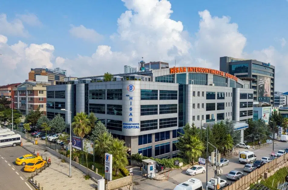

Hisar Intercontinental Hospital
Saray Mah. Siteyolu Cad. No:7, Umraniye, 34768, Istanbul, Turkey
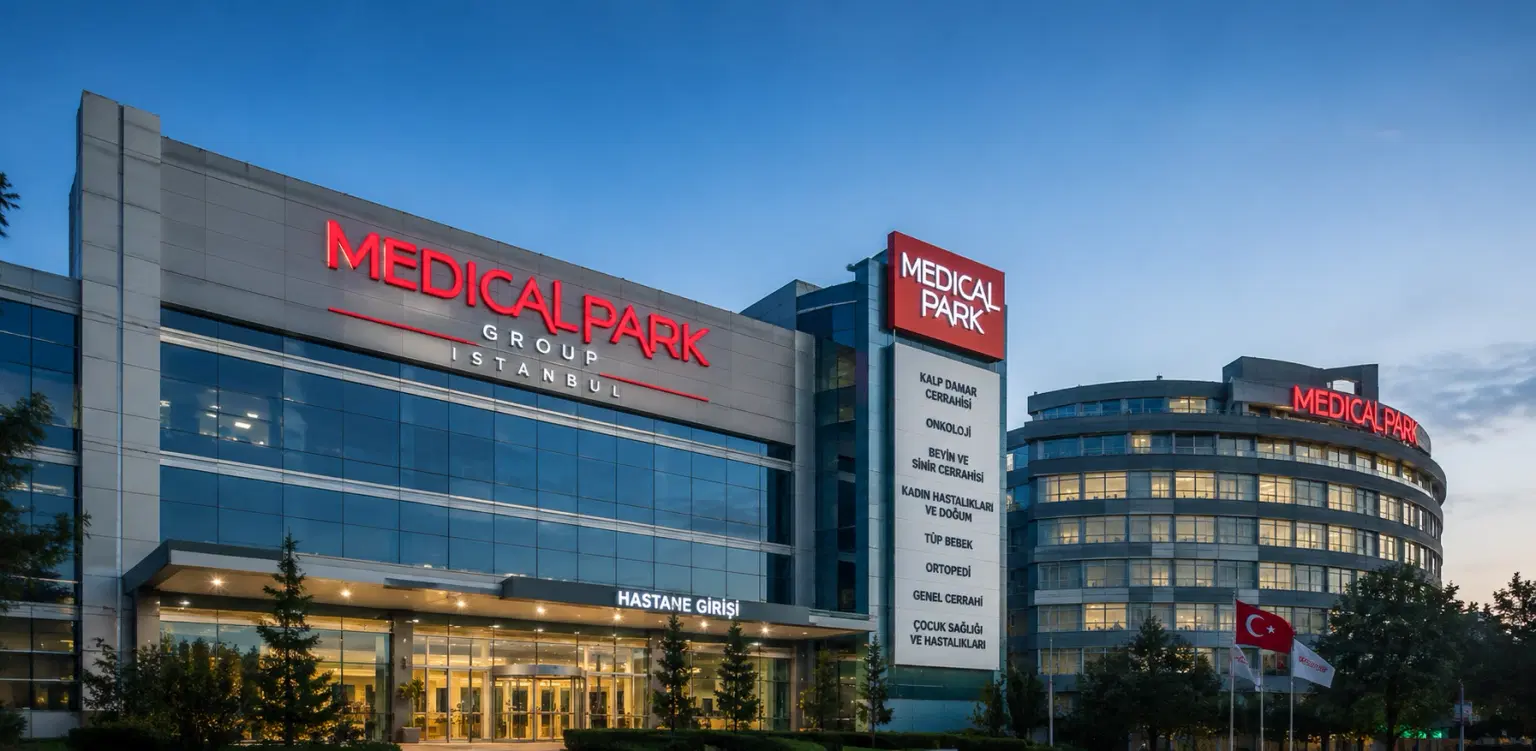
Medical Park Group, Istanbul
Fahrettin Kerim Gokay Cad. Tıbbiye Cd., Kadikoy, Istanbul, Turkey
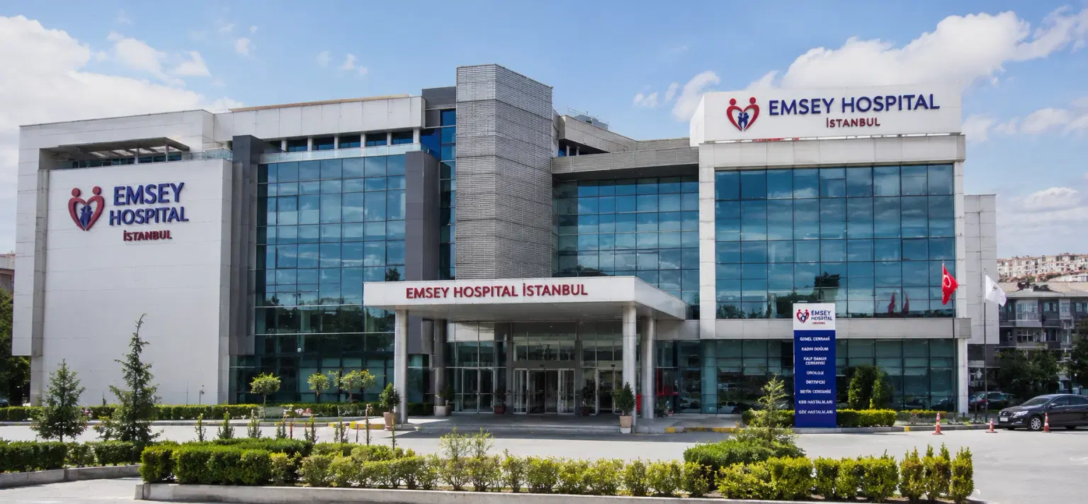
Emsey Hospital, Pendik, Istanbul
Çamlık, Selçuklu Cd. No:22, 34912 Pendik/İstanbul, Türkiye
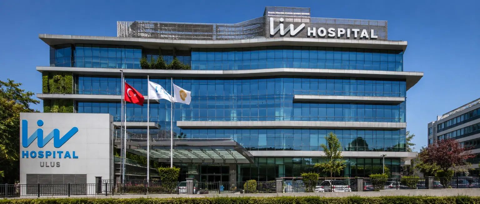
LIV Hospital, Istanbul
How DivinHeal Helps
We simplify your medical journey by providing comprehensive support and access to world-class healthcare.
Expert Specialist Matching
Connecting you with the world's top-rated medical experts.
Accredited Hospital Network
Access to JCI & NABH certified healthcare facilities.
Complete Travel Coordination
Hassle-free visa, stay, and local transport assistance.
24/7 Personal Care
Dedicated patient advisors supporting you at every step.
Journey Guidance
Full guidance from start to end of the patient treatment journey.
Expert Specialist Matching
Connecting you with the world's top-rated medical experts.
Everything you
need to know today
Browse through these common inquiries to better understand our patient-focused medical platform.
Yes, we work with a variety of insurance providers. Contact our team to verify your coverage.
Yes, we provide secure online consultations with experienced specialists.
Our care coordinators help match you with the most suitable specialist.
Absolutely. Your medical information is protected according to healthcare privacy standards.
Look at six things: accreditation (JCI or NABH), specialty depth, doctor credentials and experience, procedure-specific success rates, international patient support, and technology. DivinHeal's AI-driven matching evaluates every hospital in our accredited partner network on these dimensions and shortlists the best-fit options for your condition, budget, and country.
JCI (Joint Commission International) is the US-based global gold standard for hospital quality, recognised worldwide. NABH is India's national accreditation — accredited by ISQua, the same body that accredits JCI. Both signal independently verified safety and quality. Most of India's leading hospitals hold both.
Yes. All three welcome international patients through structured medical visa programs. India is the most established, treating patients from Africa, the Middle East, and South Asia at 60–80% lower cost. Thailand leads in cosmetic and dental care. The UAE is emerging in oncology and reproductive medicine.
Most patients save 50–80% on treatment costs. Heart bypass costs US $7,000–9,000 in India compared to $70,000–150,000 in the US. IVF costs $3,000–4,500 compared to $12,000–20,000 in the UK. Even after flights, visa, and accommodation, total savings remain 60–70%.
DivinHeal manages your entire non-medical journey: visa invitation letters, medical visa guidance, doctor appointments, teleconsultations, airport pickup, hospital-vetted accommodation for you and your attendant, language interpreters, local transport, cuisine preferences, and post-treatment follow-up — one dedicated coordinator from first enquiry to final follow-up.
You need a valid passport (6+ months validity), a medical visa (M-Visa for India — DivinHeal provides the hospital invitation letter), return flight tickets, recent medical reports and a doctor's referral, current prescription list, and proof of financial means. Any accompanying attendant needs their own passport and MX-Visa.
Still have more questions?
Book a call with our friendly team to learn how DivineHeal simplifies your healthcare journey.