Centres Of Excellence
Our Centres of Excellence bring together multidisciplinary teams to deliver precise diagnosis, advanced treatments, and superior outcomes across a wide spectrum of medical specialties.

OVERVIEW
Gastroenterology Treatment: Comprehensive Care for Digestive Health
Gastroenterology treatment focuses on the intricate health of the digestive system. At DivinHeal, we connect you with leading gastroenterologists and state-of-the-art facilities across India, offering a spectrum of care from diagnostic endoscopy to advanced surgical interventions. Our goal is to provide effective, compassionate, and affordable Gastroenterology treatment, ensuring optimal outcomes and a swift return to health for conditions ranging from common digestive complaints to complex gastrointestinal diseases.
PROCEDURE
Gastroenterology procedures range from non-invasive diagnostic tests to complex surgical interventions. Diagnostic procedures often include upper endoscopy (EGD), colonoscopy, sigmoidoscopy, capsule endoscopy, endoscopic ultrasound (EUS), and endoscopic retrograde cholangiopancreatography (ERCP) to visualize and sample tissues within the digestive tract. Therapeutic endoscopic procedures can involve polyp removal, stricture dilation, stent placement, or bleeding control. Medical management typically includes pharmacotherapy with anti-inflammatory drugs, immunosuppressants, biologics, or acid suppressants. Surgical interventions may range from minimally invasive laparoscopic procedures (e.g., cholecystectomy, appendectomy, anti-reflux surgery) to open abdominal surgeries for complex conditions like large tumor resections, bowel resections for IBD, or liver and pancreatic surgeries. For gastrointestinal cancers, Gastroenterology chemotherapy and radiation therapy, alongside Gastroenterology targeted therapy, may be integral parts of a multimodal treatment plan, aiming for disease control and improved prognosis.
BENEFITS
Benefits of Advanced Gastroenterology Treatment
Improved Digestive Function
Effective management and resolution of conditions leading to better digestion and nutrient absorption.
Pain & Symptom Relief
Alleviation of chronic abdominal pain, discomfort, bloating, and other debilitating GI symptoms.
Enhanced Quality of Life
Freedom from daily digestive struggles, allowing patients to enjoy food and activities without fear or discomfort.
Prevention of Complications
Early diagnosis and intervention can prevent progression of diseases, reduce risk of cancer, and avoid emergency situations.
Access to Innovative Therapies
Benefit from the latest advancements in medical, endoscopic, and surgical Gastroenterology diagnosis and therapy options.
RECOVERY
Gastroenterology Recovery Time and Tips
The Gastroenterology recovery time varies significantly depending on the specific procedure performed and the patient's overall health. Simple diagnostic endoscopies might involve only a few hours of recovery, while complex abdominal surgeries could require several weeks to months for full recuperation.
Post-Procedure Care
Adhere strictly to your physician's instructions regarding diet, medication, and activity levels. This often includes a gradual reintroduction of foods, pain management, and avoiding strenuous activities.
Lifestyle Modifications
Many GI conditions benefit from long-term lifestyle changes, including a balanced diet, regular exercise, stress management, and adequate hydration.
Follow-up Appointments
Regular follow-up with your gastroenterologist is crucial to monitor progress, adjust medications, and address any lingering concerns. DivinHeal assists in coordinating these appointments.
Emotional Support
Coping with chronic digestive conditions can be challenging. We encourage seeking support from family, friends, or support groups, and can help connect you with counseling services if needed for comprehensive Gastroenterology recovery.
WHAT WE TREAT
Conditions Treated in Gastroenterology
Inflammatory Bowel Disease (IBD)
Including Crohn's Disease and Ulcerative Colitis, managed through medication, dietary adjustments, and sometimes surgery.
Gastroesophageal Reflux Disease (GERD)
Treatments range from lifestyle changes and medication to advanced endoscopic or surgical interventions.
Peptic Ulcer Disease
Management of stomach and duodenal ulcers caused by H. pylori infection or NSAID use.
Liver Diseases
Such as hepatitis (viral, autoimmune), fatty liver disease, cirrhosis, and liver cancer, requiring specialized medical and interventional care.
Pancreatic Disorders
Including pancreatitis (acute and chronic), pancreatic cysts, and pancreatic cancer, often involving complex diagnosis and multidisciplinary treatment.
Gallstone Disease
Medical management or surgical removal of the gallbladder (cholecystectomy).
Celiac Disease & Malabsorption Syndromes
Dietary management and medical therapy to improve nutrient absorption.
Colorectal Polyps & GI Cancers
Early detection, endoscopic removal of polyps, and comprehensive Gastroenterology chemotherapy and radiation therapy, surgery, and Gastroenterology targeted therapy for various gastrointestinal cancers (esophageal, stomach, colorectal, liver, pancreatic).
Irritable Bowel Syndrome (IBS)
Symptom management through diet, lifestyle, and medication.
PREPARATION
Preparation for Gastroenterology treatment is highly individualized. For endoscopic procedures like colonoscopy, bowel preparation with laxatives is necessary. For upper endoscopies, an overnight fast is required. Prior to any surgical procedure, patients typically undergo comprehensive pre-operative assessments, including blood tests, imaging scans, and cardiac evaluations. Medication review is crucial; patients may be advised to stop blood thinners or certain diabetes medications before some procedures. Detailed instructions regarding diet, fasting, and medication adjustments will be provided by your medical team well in advance to ensure optimal safety and efficacy.
RISKS
While Gastroenterology treatments are generally safe, potential risks exist, varying by procedure. Common risks associated with endoscopic procedures include bleeding, infection, and rarely, perforation of the digestive tract. Surgical risks can include bleeding, infection, adverse reactions to anesthesia, wound complications, and damage to surrounding organs. For medical therapies, side effects can range from mild (nausea, fatigue) to more severe allergic reactions or immunosuppression. For treatments involving Gastroenterology chemotherapy and radiation therapy, common risks include hair loss, fatigue, nausea, and increased infection risk. Your medical team will discuss specific risks relevant to your chosen Gastroenterology diagnosis and therapy options and take all precautions to minimize them.
JOURNEY
Your Gastroenterology Treatment Journey with DivinHeal
Initial Consultation & Diagnosis
Your journey begins with a thorough evaluation by a top gastroenterologist. This involves a detailed medical history, physical examination, and advanced diagnostic tests such as endoscopy, colonoscopy, capsule endoscopy, MRI, CT scans, or specialized blood tests to accurately pinpoint the cause of your digestive issues. DivinHeal facilitates seamless appointments and secure transfer of medical records.
Personalized Treatment Planning
Based on your diagnosis, a multidisciplinary team collaborates to devise a personalized Gastroenterology diagnosis and therapy options plan. This may include medical management with medication, advanced endoscopic procedures, or surgical intervention. Your care coordinator ensures you understand every aspect of your treatment.
Treatment & Hospital Stay
Whether it's a medical regimen, an outpatient endoscopic procedure, or inpatient surgery, you'll receive care at one of India's best hospitals for Gastroenterology. Our partner hospitals boast cutting-edge technology and highly experienced medical staff, ensuring safety and efficacy. DivinHeal provides end-to-end support, from visa and travel arrangements to hospital admission and discharge.
Recovery & Follow-up
Post-treatment, our team assists with your Gastroenterology recovery time and tips. This includes coordinating follow-up appointments, providing guidance on diet and lifestyle modifications, and arranging for any necessary rehabilitation or ongoing medical management. We ensure a smooth transition back to your daily life, emphasizing long-term wellness.
OUTCOMES
Expected outcomes include significant relief from digestive symptoms, improved organ function, and a better overall quality of life. Long-term prognosis often involves successful management or resolution of conditions, enabling patients to return to normal activities with enhanced well-being.
Related Links
Doctors For Treatment in Similar Locations
- Best Gastroenterology Doctors in Bengaluru
- Best Gastroenterology Doctors in Chennai
- Best Gastroenterology Doctors in Gurugram
- Best Gastroenterology Doctors in Hyderabad
- Best Gastroenterology Doctors in India
- Best Gastroenterology Doctors in Mumbai
- Best Gastroenterology Doctors in New Delhi
- Best Gastroenterology Doctors in Pune
Best Hospital Near by for treatment
- Best Gastroenterology Hospitals in Bengaluru
- Best Gastroenterology Hospitals in Chennai
- Best Gastroenterology Hospitals in Gurugram
- Best Gastroenterology Hospitals in Hyderabad
- Best Gastroenterology Hospitals in India
- Best Gastroenterology Hospitals in Mumbai
- Best Gastroenterology Hospitals in New Delhi
- Best Gastroenterology Hospitals in Pune
Related Treatments

IBD (Crohn's / Ulcerative Colitis) Management
Treatment Price
$0.00 USDNo overview available
Treatment Overview
No overview available
Procedures
No procedure details available
Benefits
No benefits information available
Recovery Information
No recovery information available
Bariatric Endoscopy (ESG, Balloon)
Treatment Price
$0.00 USDNo overview available
Treatment Overview
No overview available
Procedures
No procedure details available
Benefits
No benefits information available
Recovery Information
No recovery information available
GERD / Reflux Surgery (Fundoplication)
Treatment Price
$0.00 USDNo overview available
Treatment Overview
No overview available
Procedures
No procedure details available
Benefits
No benefits information available
Recovery Information
No recovery information available
Pancreatitis Management
Treatment Price
$0.00 USDNo overview available
Treatment Overview
No overview available
Procedures
No procedure details available
Benefits
No benefits information available
Recovery Information
No recovery information available
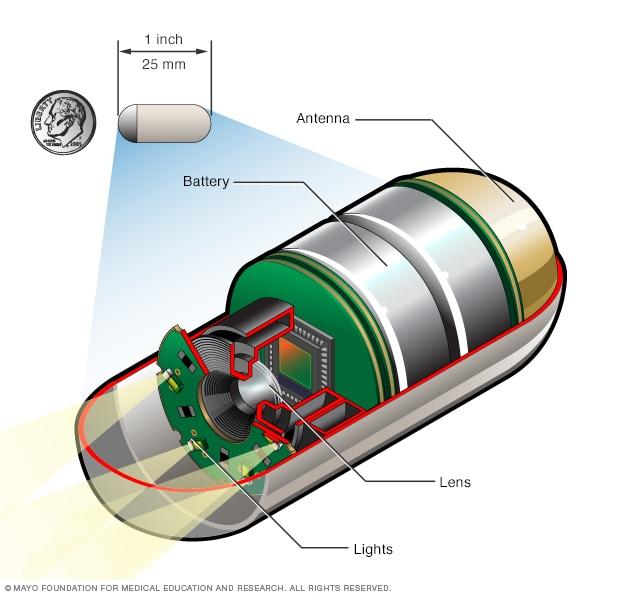
Capsule Endoscopy
Treatment Price
$900.00 USDCapsule Endoscopy provides detailed images of the small bowel, crucial for diagnosing conditions like unexplained gastrointestinal bleeding, Crohn's disease, celiac disease, and small bowel tumors. It offers a comfortable, radiation-free alternative to explore difficult-to-reach areas of the digestive tract.
Treatment Overview
Capsule Endoscopy provides detailed images of the small bowel, crucial for diagnosing conditions like unexplained gastrointestinal bleeding, Crohn's disease, celiac disease, and small bowel tumors. It offers a comfortable, radiation-free alternative to explore difficult-to-reach areas of the digestive tract.
Procedures
The Capsule Endoscopy procedure is remarkably simple for the patient.1. **Preparation**: Patients typically fast for 8-12 hours prior to the procedure. Clear liquids might be allowed up to two hours before.2. **Swallowing the Capsule**: The patient swallows a vitamin-sized capsule, usually with a small sip of water. This capsule contains a miniature camera, light source, battery, and transmitter.3. **Wearing the Data Recorder**: A belt with sensors and a data recorder is fitted around the patient's waist. This device wirelessly receives and stores the images transmitted by the capsule.4. **Image Capture**: As the capsule travels through the small intestine, it continuously captures images (typically 2-6 frames per second) for about 8-12 hours.5. **Normal Activity**: Patients can usually resume light activities during the recording period, following specific instructions regarding eating, drinking, and physical exertion.6. **Completion**: After the recording period, the patient returns the data recorder to the clinic.7. **Analysis**: The stored images are downloaded to a computer and compiled into a video for review by a gastroenterologist, who analyzes the footage for abnormalities.8. **Natural Passage**: The capsule passes naturally and painlessly through the digestive system and is excreted in a bowel movement, typically within 24-72 hours.
Benefits
<h2>Key Benefits of Capsule Endoscopy</h2><ul><li><h3>Non-Invasive and Painless:</h3> Unlike traditional endoscopic procedures, Capsule Endoscopy requires no sedation, incisions, or discomfort. Patients simply swallow a pill-sized camera.</li><li><h3>Comprehensive Small Bowel Visualization:</h3> It offers an unparalleled view of the entire small intestine, an area difficult to reach with conventional endoscopes, crucial for detecting obscure conditions.</li><li><h3>High Diagnostic Accuracy:</h3> Highly effective in identifying sources of unexplained gastrointestinal bleeding, Crohn's disease, celiac disease, and small bowel tumors.</li><li><h3>Radiation-Free:</h3> An excellent alternative to imaging techniques that involve radiation exposure, such as CT scans.</li><li><h3>Patient Comfort and Convenience:</h3> Patients can typically go about their daily routine during the procedure, making it far less disruptive than other diagnostic tests.</li><li><h3>Early Detection:</h3> Aids in the early detection of various gastrointestinal conditions, allowing for timely intervention and improved outcomes.</li></ul>
Recovery Information
<h2>Recovery and Post-Procedure Care for Capsule Endoscopy</h2><p>Recovery from a Capsule Endoscopy is exceptionally straightforward and typically involves no downtime. Since the procedure is non-invasive and does not require sedation, you can usually resume your normal activities immediately after swallowing the capsule.</p><h3>What to Expect Immediately After:</h3><ul><li>You will wear a data recorder on a belt for approximately 8 hours, capturing images from the capsule as it travels through your digestive system.</li><li>You can usually drink clear liquids after two hours and have a light meal four hours after swallowing the capsule, unless otherwise advised by your doctor.</li><li>Avoid strenuous physical activity or excessive bending during the recording period, as this can interfere with image transmission.</li></ul><h3>Capsule Passage:</h3><ul><li>The capsule is disposable and designed to pass naturally and painlessly out of your body with a bowel movement, typically within 24-72 hours. It does not need to be retrieved or returned.</li></ul><h3>Receiving Results:</h3><ul><li>Once you return the data recorder, the images are downloaded and carefully reviewed by a gastroenterologist. This process can take a few days.</li><li>Your doctor will schedule a follow-up appointment to discuss the findings and determine any necessary next steps, such as further diagnostic tests or treatment plans.</li></ul><p>DivinHeal ensures you are guided through every step, from preparation to understanding your results, connecting you with specialists who will provide clear, compassionate explanations.</p>

Colonoscopy with Polypectomy
Treatment Price
$1000.00 USDThis procedure involves inserting a flexible tube with a camera (colonoscope) to visualize the colon's inner lining. If polyps are found, they are removed during the same procedure (polypectomy). It's a cornerstone for early detection and prevention of colorectal cancer, offering both diagnostic clarity and immediate therapeutic intervention.
Treatment Overview
This procedure involves inserting a flexible tube with a camera (colonoscope) to visualize the colon's inner lining. If polyps are found, they are removed during the same procedure (polypectomy). It's a cornerstone for early detection and prevention of colorectal cancer, offering both diagnostic clarity and immediate therapeutic intervention.
Procedures
The Colonoscopy with Polypectomy procedure typically involves several key steps: 1. **Sedation:** Upon arrival, you will be given a sedative and pain medication intravenously to ensure comfort and relaxation throughout the procedure. 2. **Insertion of Colonoscope:** Once sedated, the doctor gently inserts the colonoscope into the rectum and advances it through the entire large intestine to the cecum (the beginning of the colon). 3. **Visualization:** The colonoscope transmits images to a video monitor, allowing the gastroenterologist to thoroughly examine the colon lining for polyps, inflammation, bleeding, or other abnormalities. Air is often gently pumped into the colon to expand it, providing a clearer view. 4. **Polypectomy (if necessary):** If polyps are identified, they are removed using specialized instruments passed through the colonoscope. Small polyps may be removed with biopsy forceps, while larger ones are typically removed with a snare (a thin wire loop that is tightened around the base of the polyp and then cauterized to seal the blood vessels). 5. **Biopsy (if necessary):** If other suspicious areas are found, small tissue samples (biopsies) may be taken for further laboratory analysis. 6. **Withdrawal of Colonoscope:** After the examination and any necessary interventions, the colonoscope is slowly and carefully withdrawn. The entire procedure usually takes between 30 to 60 minutes, depending on the findings and interventions required.
Benefits
<h2>Key Benefits of Colonoscopy with Polypectomy</h2><ul><li><h3>Colorectal Cancer Prevention</h3><p>The most significant benefit is the ability to remove precancerous polyps, effectively preventing the development of colorectal cancer.</p></li><li><h3>Early Detection of Cancer</h3><p>Allows for the detection of colorectal cancer at its earliest, most treatable stages, leading to higher <a href="Colonoscopy-with-Polypectomy-success-rate">Colonoscopy with Polypectomy success rates</a>.</p></li><li><h3>Diagnostic Accuracy</h3><p>Provides a direct visual examination of the entire colon, offering superior diagnostic accuracy compared to other screening methods.</p></li><li><h3>Relief of Symptoms</h3><p>Can identify and sometimes treat sources of symptoms like rectal bleeding, abdominal pain, or changes in bowel habits.</p></li><li><h3>Minimally Invasive Treatment</h3><p>Polypectomy is performed endoscopically, avoiding open surgery and its associated longer recovery times.</p></li></ul>
Recovery Information
<h2><a href="Colonoscopy-with-Polypectomy-recovery-time-and-tips">Recovery Time and Tips</a> After Colonoscopy with Polypectomy</h2><h3>Immediate Post-Procedure</h3><p>Most patients recover quickly from a Colonoscopy with Polypectomy. You will typically spend an hour or two in a recovery area while the sedation wears off. You might experience mild cramping, bloating, or gas, which usually resolves within a few hours. Due to the sedation, it's mandatory to have someone drive you home.</p><h3>First 24 Hours</h3><p>Avoid driving, operating heavy machinery, making important decisions, and consuming alcohol for at least 24 hours post-procedure. You can generally resume light activities and a normal diet. Some may be advised to avoid fibrous foods initially if multiple or large polyps were removed.</p><h3>Long-Term Recovery & Follow-up</h3><p>Full recovery is usually within 24-48 hours. Your gastroenterologist will discuss the findings, including the pathology results of any removed polyps, and provide recommendations for your next screening or follow-up. Adhering to these recommendations is crucial for continued colorectal health.</p><p>DivinHeal ensures you receive clear, comprehensive post-procedure instructions and facilitates any necessary follow-up consultations with your <a href="Top-gastroenterologists-for-Colonoscopy-with-Polypectomy">top gastroenterologists for Colonoscopy with Polypectomy</a>.</p>

Liver Fibroscan
Treatment Price
$300.00 USDLiver Fibroscan utilizes ultrasound technology to measure the elasticity of liver tissue, providing a reliable indicator of fibrosis, and an attenuation parameter (CAP) to quantify steatosis. This procedure helps diagnose conditions like fatty liver disease (NAFLD/NASH), hepatitis, and cirrhosis, guiding timely intervention and treatment strategies.
Treatment Overview
Liver Fibroscan utilizes ultrasound technology to measure the elasticity of liver tissue, providing a reliable indicator of fibrosis, and an attenuation parameter (CAP) to quantify steatosis. This procedure helps diagnose conditions like fatty liver disease (NAFLD/NASH), hepatitis, and cirrhosis, guiding timely intervention and treatment strategies.
Procedures
A Liver Fibroscan is performed with the patient lying on their back with their right arm raised above their head. A trained technician applies a water-based gel to the skin over the liver area (between the ribs). A probe, similar to an ultrasound probe, is placed on the skin and delivers gentle vibrations. These vibrations create a sheer wave that travels through the liver, and the device measures the speed of this wave to determine liver stiffness. The procedure takes about 5-10 minutes, involving typically 10 successful measurements to ensure accuracy.
Benefits
The primary benefits of Liver Fibroscan include its non-invasive nature, making it a safe and comfortable procedure. It provides rapid, quantitative results for liver stiffness (fibrosis) and fat content (steatosis), aiding in early diagnosis and monitoring of liver diseases. It reduces the need for painful and risky liver biopsies, offering a reliable tool for tracking disease progression and assessing treatment efficacy over time.
Recovery Information
Recovery from a Liver Fibroscan is immediate. As a non-invasive procedure, there is no downtime, anesthesia, or special post-procedure care required. Patients can resume their normal activities immediately after the scan. The results are typically available shortly after the procedure, allowing for prompt discussion with your physician.
Gallbladder Surgery (Laparoscopic Cholecystectomy)
Treatment Price
$3500.00 USDUnderstanding Gallbladder Surgery (Laparoscopic Cholecystectomy)
The primary goal of Gallbladder Surgery (Laparoscopic Cholecystectomy) is to remove the gallbladder, a small organ responsible for storing bile, when it becomes a source of pain, infection, or other complications, most commonly due to gallstones. The laparoscopic technique involves making several small incisions instead of one large one, leading to less pain, smaller scars, and a quicker recovery compared to traditional open surgery. DivinHeal connects you with top surgeons and accredited facilities ensuring high-quality, safe, and effective treatment outcomes.
Treatment Overview
<h2>Understanding Gallbladder Surgery (Laparoscopic Cholecystectomy)</h2><p>The primary goal of Gallbladder Surgery (Laparoscopic Cholecystectomy) is to remove the gallbladder, a small organ responsible for storing bile, when it becomes a source of pain, infection, or other complications, most commonly due to gallstones. The laparoscopic technique involves making several small incisions instead of one large one, leading to less pain, smaller scars, and a quicker recovery compared to traditional open surgery. DivinHeal connects you with top surgeons and accredited facilities ensuring high-quality, safe, and effective treatment outcomes.</p>
Procedures
Laparoscopic Cholecystectomy is performed under general anesthesia. The surgeon makes 3-4 small incisions (0.5-1 cm) in the abdomen. Carbon dioxide gas is inflated into the abdominal cavity to create space and better visibility. A laparoscope, a thin tube with a camera, is inserted through one incision, displaying magnified images on a monitor. Surgical instruments are inserted through other incisions. The gallbladder is carefully dissected from the liver and surrounding structures, the cystic duct and artery are clipped and cut, and the gallbladder is then removed through one of the incisions. Finally, the carbon dioxide gas is released, and the incisions are closed with sutures or surgical glue.
Benefits
<h2>Key Benefits of Laparoscopic Cholecystectomy</h2><ul><li><h3>Effective Relief from Symptoms:</h3><p>Provides definitive relief from the excruciating pain, nausea, and digestive issues caused by gallstones and cholecystitis.</p><li><h3>Minimally Invasive:</h3><p>Smaller incisions lead to less pain, reduced scarring, and a lower risk of wound complications compared to open surgery.</p><li><h3>Faster Recovery Time:</h3><p>Patients typically return to light activities within a few days and full activities within 1-2 weeks, significantly faster than traditional methods.</p><li><h3>Shorter Hospital Stay:</h3><p>Most patients are discharged within 24-48 hours, reducing the overall healthcare cost and inconvenience.</p><li><h3>High Success Rate:</h3><p>Laparoscopic Cholecystectomy has a very high success rate in resolving gallbladder-related issues permanently.</p><li><h3>Improved Quality of Life:</h3><p>Freedom from recurrent pain and digestive problems allows for a significant improvement in daily living and dietary freedom (with some initial adjustments).</p></li></li></li></li></li></li></ul>
Recovery Information
<h2>Gallbladder Surgery (Laparoscopic Cholecystectomy) Recovery Time and Tips</h2><ul><li><h3>Immediate Post-operative Period (1-2 Days):</h3><p>You will typically stay in the hospital for 1-2 days. Pain medication will be administered to manage discomfort. You'll be encouraged to walk soon after surgery to aid recovery and prevent complications like blood clots.</p><li><h3>First Week Home:</h3><p>Expect some mild pain or discomfort around the incision sites and possibly in your shoulder (due to residual gas from surgery). Rest is important, but gentle activity like short walks is beneficial. Avoid heavy lifting and strenuous exercise. Follow a bland, low-fat diet initially and gradually reintroduce foods.</p><li><h3>Weeks 2-4:</h3><p>Most patients can return to normal, non-strenuous activities, including work (if not physically demanding). Incision sites should be healing well. Continue to avoid heavy lifting. You may experience some changes in bowel habits initially as your body adjusts to the absence of the gallbladder.</p><li><h3>Long-Term Recovery and Lifestyle:</h3><p>Full recovery, where you feel completely back to normal, usually takes about 4-6 weeks. Most people live a normal life without a gallbladder, with no significant dietary restrictions long-term. However, some may find they tolerate high-fat meals less well. DivinHeal provides comprehensive post-operative guidance and connects you with dieticians if needed.</p></li></li></li></li></ul>
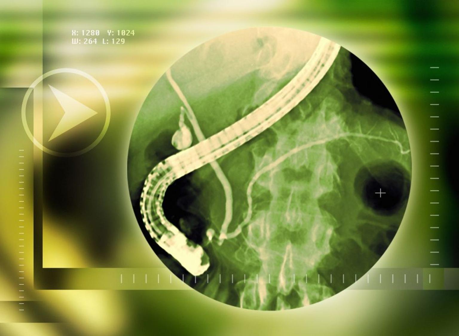
ERCP (Endoscopic Retrograde Cholangiopancreatography)
Treatment Price
$2000.00 USDERCP is primarily a therapeutic endoscopic procedure, though it can also be diagnostic. Its main goals include removing gallstones from bile ducts, opening narrowed ducts (strictures), taking biopsies of suspicious growths, and draining blocked bile ducts. It involves inserting a flexible tube through the mouth, stomach, and duodenum, guided by fluoroscopy, to access the bile and pancreatic ducts.
Treatment Overview
ERCP is primarily a therapeutic endoscopic procedure, though it can also be diagnostic. Its main goals include removing gallstones from bile ducts, opening narrowed ducts (strictures), taking biopsies of suspicious growths, and draining blocked bile ducts. It involves inserting a flexible tube through the mouth, stomach, and duodenum, guided by fluoroscopy, to access the bile and pancreatic ducts.
Procedures
The ERCP procedure involves several key steps. First, the patient is given sedation or general anesthesia. The gastroenterologist then inserts a thin, flexible endoscope through the mouth, down the esophagus, stomach, and into the duodenum, reaching the papilla of Vater. A small catheter is then advanced through the endoscope into the bile or pancreatic duct. Contrast dye is injected, and X-ray images (fluoroscopy) are taken to visualize the ducts and identify any abnormalities like stones or strictures. If issues are found, therapeutic interventions can be performed, such as sphincterotomy (making a small cut in the papilla), stone removal using a basket or balloon, or stent placement to open a blocked duct. Biopsies can also be taken. After the interventions, the endoscope is slowly withdrawn.
Benefits
<h2>Benefits of ERCP Treatment</h2><ul><li><strong>Minimally Invasive:</strong> Avoids open surgery, leading to smaller incisions, less pain, and quicker recovery.</li><li><strong>Diagnostic & Therapeutic:</strong> Can both identify and treat issues in a single procedure.</li><li><strong>Effective Stone Removal:</strong> Highly effective in clearing bile duct stones that cause jaundice or pancreatitis.</li><li><strong>Stricture Relief:</strong> Opens narrowed ducts, restoring normal bile or pancreatic fluid flow.</li><li><strong>Biopsy Capabilities:</strong> Allows for tissue sampling to accurately diagnose conditions, including cancers.</li><li><strong>Reduces Symptoms:</strong> Rapidly alleviates pain, jaundice, and other debilitating symptoms caused by blockages.</li><li><strong>High Success Rate:</strong> When performed by experienced specialists, ERCP has a high success rate for its intended purposes.</li></ul>
Recovery Information
<h2>ERCP Recovery Time and Tips</h2><p>Recovery after an ERCP is generally swift, with most patients returning home on the same day or staying overnight for observation. The immediate recovery involves:</p><ul><li><strong>Post-Procedure Monitoring:</strong> Patients are monitored in a recovery area for a few hours until the sedation wears off. Vital signs are checked, and for signs of complications like pancreatitis.</li><li><strong>Diet:</strong> Most patients can resume a light diet a few hours after the procedure, once the gag reflex returns. Your doctor will provide specific dietary instructions.</li><li><strong>Discomfort:</strong> Mild sore throat, bloating, or gas are common and usually resolve within a day or two. Pain medication can be prescribed if needed.</li><li><strong>Rest:</strong> It's advisable to rest for the remainder of the day after the procedure and avoid strenuous activities.</li></ul><h3>Long-Term Wellness</h3><p>For most, there are no significant long-term recovery issues specific to the ERCP itself. However, follow-up care for the underlying condition (e.g., gallstones, pancreatitis) will be crucial. Your doctor may recommend:</p><ul><li>Lifestyle modifications or dietary changes if the underlying cause was related to diet.</li><li>Follow-up imaging or endoscopic procedures depending on the diagnosis.</li><li>Continued management for chronic conditions like recurrent pancreatitis.</li></ul><p>DivinHeal's care coordinators ensure you have clear post-procedure instructions and connect you with ongoing support as needed.</p>
Colonoscopy (Diagnostic)
Treatment Price
$450.00 USDColonoscopy (Diagnostic) aims to visualize the inner lining of the colon and rectum, identifying potential issues such as polyps (precancerous growths), inflammation, ulcers, or tumors. During the procedure, small tissue samples (biopsies) can be taken, and polyps can be removed (polypectomy) to prevent the development of colorectal cancer. It's a gold standard for colorectal cancer screening and diagnosis.
Treatment Overview
Colonoscopy (Diagnostic) aims to visualize the inner lining of the colon and rectum, identifying potential issues such as polyps (precancerous growths), inflammation, ulcers, or tumors. During the procedure, small tissue samples (biopsies) can be taken, and polyps can be removed (polypectomy) to prevent the development of colorectal cancer. It's a gold standard for colorectal cancer screening and diagnosis.
Procedures
A Colonoscopy (Diagnostic) involves the patient lying on their side while a thin, flexible tube called a colonoscope is gently inserted into the rectum and advanced through the colon. The colonoscope has a light and a tiny camera that transmits images to a monitor, allowing the gastroenterologist to thoroughly examine the colon lining. Air is often inflated into the colon to improve visibility. If polyps or suspicious areas are found, special instruments passed through the scope can remove polyps or take biopsies. The procedure typically takes 30-60 minutes.
Benefits
<ul><li><strong>Early Detection & Prevention:</strong> The primary benefit is the early detection and removal of precancerous polyps, significantly reducing the risk of colorectal cancer.</li><li><strong>Accurate Diagnosis:</strong> Provides definitive answers for unexplained bowel symptoms, guiding appropriate treatment.</li><li><strong>Minimally Invasive:</strong> A relatively safe, outpatient procedure with a quick recovery for most patients.</li><li><strong>Therapeutic Capabilities:</strong> Polyps can be removed and biopsies taken during the same procedure, avoiding additional interventions.</li><li><strong>Peace of Mind:</strong> Timely screening offers reassurance and contributes to long-term health.</li></ul>
Recovery Information
Recovery from a Colonoscopy (Diagnostic) is typically quick. Patients usually spend 1-2 hours in a recovery area post-procedure as the effects of sedation wear off. You may experience some mild bloating or gas for a few hours. It is advised to rest for the remainder of the day and avoid driving or operating machinery. Most individuals can resume normal activities the following day, though specific dietary recommendations might be given for a short period. DivinHeal ensures you receive clear post-procedure instructions for a smooth and safe recovery.
Liver Biopsy
Treatment Price
$600.00 USDLiver Biopsy aims to diagnose liver diseases, assess liver damage, and monitor treatment effectiveness when non-invasive tests are inconclusive. Techniques include percutaneous, transjugular, or laparoscopic approaches, chosen based on the patient's condition and the physician's assessment. DivinHeal connects you with experienced hepatologists and world-class facilities for accurate diagnosis.
Treatment Overview
Liver Biopsy aims to diagnose liver diseases, assess liver damage, and monitor treatment effectiveness when non-invasive tests are inconclusive. Techniques include percutaneous, transjugular, or laparoscopic approaches, chosen based on the patient's condition and the physician's assessment. DivinHeal connects you with experienced hepatologists and world-class facilities for accurate diagnosis.
Procedures
A Liver Biopsy typically involves the following steps: 1. Patient preparation: Fasting for several hours, blood tests to check clotting ability, and sometimes a mild sedative. 2. Positioning: The patient lies on their back with their right arm raised above their head to expose the right side of the abdomen. 3. Local Anesthesia: The skin over the biopsy site is numbed with local anesthetic. 4. Imaging Guidance: Ultrasound or CT scan is used to precisely locate the liver and guide the biopsy needle, avoiding blood vessels or other organs. 5. Needle Insertion: A specialized biopsy needle is quickly inserted through the skin and into the liver. The patient is usually asked to hold their breath briefly during insertion to minimize liver movement. 6. Sample Collection: A small tissue sample is extracted. Sometimes, multiple samples are taken. 7. Needle Removal: The needle is quickly withdrawn. 8. Pressure Application: Pressure is applied to the biopsy site to minimize bleeding, and a dressing is applied. 9. Post-Procedure Monitoring: The patient is monitored in a recovery area for several hours for any complications.
Benefits
<h2>Benefits of Undergoing a Liver Biopsy</h2><ul><li><h3>Accurate Diagnosis</h3><p>A Liver Biopsy provides definitive diagnostic information that often cannot be obtained through non-invasive tests alone. This precision is crucial for conditions like fibrosis, specific types of hepatitis, or rare liver disorders.</p></li><li><h3>Disease Staging and Prognosis</h3><p>It allows medical professionals to accurately stage the severity of liver disease, such as cirrhosis, helping to predict disease progression and inform prognosis.</p></li><li><h3>Tailored Treatment Plans</h3><p>With a precise diagnosis, doctors can formulate the most effective and personalized treatment plan, avoiding unnecessary or ineffective therapies.</p></li><li><h3>Monitoring Treatment Effectiveness</h3><p>For patients undergoing treatment for chronic liver conditions, a biopsy can help assess how well the liver is responding to therapy.</p></li><li><h3>Investigation of Liver Masses</h3><p>It's invaluable for differentiating between benign and malignant liver lesions, guiding decisions on further management.</p></li></ul>
Recovery Information
<h2>Liver Biopsy Recovery Time and Tips</h2><p>Recovery after a Liver Biopsy is generally quick, especially for percutaneous or transjugular methods, with most patients returning home on the same day. Here’s what to expect:</p><ul><li><h3>Immediate Post-Procedure</h3><p>You will be monitored for several hours in the recovery room to ensure no bleeding or complications occur. You might feel some mild pain or soreness at the biopsy site or in your right shoulder (referred pain). Pain medication will be provided if needed.</p></li><li><h3>First 24-48 Hours</h3><p>Rest is crucial. Avoid strenuous activities, heavy lifting, or vigorous exercise. Keep the biopsy site clean and dry. You will typically be advised to avoid driving for 24 hours.</p></li><li><h3>Full Recovery</h3><p>Most patients can resume normal, light activities within 1-2 days. Full recovery, including resuming strenuous exercise, is usually advised after about a week to allow the liver to heal completely. Your doctor will provide specific instructions tailored to your procedure and overall health.</p></li><li><h3>Important Recovery Tips</h3><ul><li><strong>Pain Management:</strong> Take prescribed pain relievers as directed.</li><li><strong>Activity Restrictions:</strong> Adhere strictly to your doctor's advice on physical activity.</li><li><strong>Monitoring:</strong> Watch for any signs of complications such as excessive bleeding, severe pain, fever, or difficulty breathing, and contact your doctor immediately if they occur.</li><li><strong>Hydration and Nutrition:</strong> Maintain good hydration and a balanced diet to support healing.</li></ul></li></ul>
Upper GI Endoscopy (Gastroscopy)
Treatment Price
$300.00 USDGastroscopy is a crucial diagnostic tool in gastroenterology. Its primary goal is to identify abnormalities within the upper digestive tract that cannot be seen on X-rays. It involves the use of an endoscope, allowing the physician to directly observe the mucous membranes, take tissue samples (biopsies) for further analysis, and perform minor therapeutic interventions such as polyp removal or stopping active bleeding. DivinHeal connects patients with leading gastroenterologists and state-of-the-art facilities for precise and safe endoscopic evaluations.
Treatment Overview
Gastroscopy is a crucial diagnostic tool in gastroenterology. Its primary goal is to identify abnormalities within the upper digestive tract that cannot be seen on X-rays. It involves the use of an endoscope, allowing the physician to directly observe the mucous membranes, take tissue samples (biopsies) for further analysis, and perform minor therapeutic interventions such as polyp removal or stopping active bleeding. DivinHeal connects patients with leading gastroenterologists and state-of-the-art facilities for precise and safe endoscopic evaluations.
Procedures
The Upper GI Endoscopy (Gastroscopy) procedure typically involves these steps: 1. **Preparation:** You will be asked to fast for 6-8 hours prior to the procedure to ensure an empty stomach. 2. **Sedation:** Upon arrival, an intravenous line will be started, and a sedative will be administered to help you relax and feel comfortable during the procedure. Local anesthetic spray may also be applied to your throat. 3. **Positioning:** You will lie on your left side on an examination table. 4. **Endoscope Insertion:** The gastroenterologist will gently guide the thin, flexible endoscope through your mouth, down your esophagus, into your stomach, and then into the duodenum. You may be asked to swallow during insertion. 5. **Examination:** Air is gently insufflated through the endoscope to expand the upper digestive tract, allowing for a clear view of the lining. The doctor meticulously examines the tissues for any abnormalities, taking still images or video recordings. 6. **Biopsy/Intervention (if needed):** If suspicious areas are found, tiny instruments passed through the endoscope can take tissue samples (biopsies) or perform minor therapeutic actions like polyp removal or stopping bleeding. 7. **Withdrawal:** Once the examination is complete, the endoscope is slowly and carefully withdrawn. 8. **Recovery:** You will be taken to a recovery area for monitoring until the sedation effects wear off.
Benefits
The benefits of undergoing an Upper GI Endoscopy are significant and contribute to accurate diagnosis and effective management of digestive health issues:<ul><li><strong>Precise Diagnosis:</strong> Direct visualization allows for accurate identification of ulcers, inflammation, polyps, tumors, and other abnormalities.</li><li><strong>Biopsy Capabilities:</strong> Tissue samples can be taken to test for conditions like H. pylori, Celiac disease, or to determine if a lesion is benign or malignant.</li><li><strong>Minor Therapeutic Interventions:</strong> Polyps can be removed, actively bleeding lesions can be treated, and narrowed areas (strictures) can be dilated during the same procedure.</li><li><strong>Minimally Invasive:</strong> It's a non-surgical procedure with minimal discomfort, typically performed under sedation, allowing for quick recovery.</li><li><strong>Early Detection:</strong> Can detect pre-cancerous conditions or early-stage cancers, significantly improving prognosis.</li><li><strong>Reassurance:</strong> Provides peace of mind by confirming or ruling out serious conditions, guiding appropriate treatment plans.</li></ul>
Recovery Information
Recovery from an Upper GI Endoscopy is generally quick and straightforward. You will typically be monitored for 1-2 hours in a recovery area until the effects of sedation wear off. It's common to experience a mild sore throat, bloating from the air introduced during the procedure, or a slight feeling of drowsiness. You should avoid driving, operating heavy machinery, or making important decisions for the rest of the day due to the lingering effects of sedation. Most patients can resume light activities the following day and return to their normal diet as soon as the sore throat subsides. Your doctor will provide specific instructions regarding diet, medication, and follow-up based on your individual findings.
Hernia Repair (Laparoscopic/Open)
Treatment Price
$2500.00 USDHernia Repair: Goals and Techniques
The primary goal of hernia repair is to push the protruding tissue back into place and reinforce the weakened abdominal wall to prevent recurrence. This can be achieved through:
Open Hernia Repair:
A single, larger incision is made near the hernia site. The surgeon pushes the bulge back in and often uses synthetic mesh to strengthen the area. This technique is well-established and effective.
Laparoscopic Hernia Repair:
Several small incisions are made, through which a laparoscope (a thin, lighted tube with a camera) and surgical instruments are inserted. The surgeon views the procedure on a monitor and repairs the hernia, typically using mesh, with less trauma to surrounding tissues. This often results in less pain and a faster recovery.
DivinHeal ensures access to experienced surgeons proficient in both techniques, offering personalized treatment plans based on the type of hernia, patient health, and recovery preferences.
Treatment Overview
<h2>Hernia Repair: Goals and Techniques</h2><p>The primary goal of hernia repair is to push the protruding tissue back into place and reinforce the weakened abdominal wall to prevent recurrence. This can be achieved through:</p><ul><li><h3>Open Hernia Repair:</h3><p>A single, larger incision is made near the hernia site. The surgeon pushes the bulge back in and often uses synthetic mesh to strengthen the area. This technique is well-established and effective.</p></li><li><h3>Laparoscopic Hernia Repair:</h3><p>Several small incisions are made, through which a laparoscope (a thin, lighted tube with a camera) and surgical instruments are inserted. The surgeon views the procedure on a monitor and repairs the hernia, typically using mesh, with less trauma to surrounding tissues. This often results in less pain and a faster recovery.</p></li></ul><p>DivinHeal ensures access to experienced surgeons proficient in both techniques, offering personalized treatment plans based on the type of hernia, patient health, and recovery preferences.</p>
Procedures
**Laparoscopic Hernia Repair:** The patient is given general anesthesia. The surgeon makes several small incisions in the abdomen. A laparoscope (a thin, lighted tube with a camera) is inserted through one incision, allowing the surgeon to view the hernia on a monitor. Surgical instruments are inserted through other small incisions. The protruding tissue is gently pushed back into the abdominal cavity. A synthetic mesh is then placed over the weakened area to reinforce the abdominal wall and stapled or glued into place. Incisions are closed with sutures or surgical tape. **Open Hernia Repair:** The patient is given general or local anesthesia with sedation. The surgeon makes a single incision, usually 3-6 inches long, near the hernia site. The layers of tissue are carefully separated to expose the hernia sac. The protruding tissue is gently pushed back into the abdomen. The weakened muscle wall is then repaired, often by suturing the edges of the healthy muscle tissue together. In most cases, a synthetic mesh patch is sewn over the weakened area to provide additional strength and prevent recurrence. The incision is then closed in layers.
Benefits
<h2>Benefits of Hernia Repair</h2><p>Undergoing hernia repair surgery offers numerous significant benefits, improving quality of life and preventing serious complications:</p><ul><li><h3>Pain Relief:</h3><p>Eliminates chronic pain and discomfort caused by the hernia, allowing for normal daily activities.</p></li><li><h3>Prevention of Complications:</h3><p>Crucially prevents serious issues like incarceration (trapping of tissue) and strangulation (loss of blood supply), which are life-threatening emergencies.</p></li><li><h3>Restored Abdominal Integrity:</h3><p>Repairs the weakened muscle wall, restoring normal anatomy and strength to the affected area.</p></li><li><h3>Improved Quality of Life:</h3><p>Patients can resume physical activities, exercise, and daily routines without fear of pain or worsening symptoms.</p></li><li><h3>Minimally Invasive Options:</h3><p>Laparoscopic repair often leads to smaller incisions, reduced post-operative pain, shorter hospital stays, and a faster return to work and normal activities compared to open surgery.</p></li><li><h3>High Success Rate:</h3><p>Hernia repair is a very common procedure with an excellent success rate, especially when performed by experienced surgeons in well-equipped facilities.</p></li></ul>
Recovery Information
<h2>Recovery and Post-Hernia Repair Care</h2><p>Recovery from hernia repair surgery varies depending on the type of procedure (laparoscopic vs. open) and individual factors. DivinHeal provides comprehensive guidance for a smooth recovery.</p><ul><li><h3>Immediate Post-operative Period (1-3 days):</h3><p>You may experience some pain, swelling, and bruising at the incision sites. Pain medication will be prescribed. For laparoscopic repair, pain is typically less severe. Most patients are discharged within 1-2 days.</p></li><li><h3>First 1-2 Weeks:</h3><p>Avoid heavy lifting, strenuous activities, and straining. Walking is encouraged to promote circulation. Keep incision sites clean and dry. You can usually return to light daily activities and desk work. For open repair, full recovery might take slightly longer.</p></li><li><h3>Weeks 3-6 and Beyond:</h3><p>Gradually increase activity levels as tolerated, but continue to avoid heavy lifting. Most patients can resume normal activities, including moderate exercise, by 4-6 weeks post-surgery. Full recovery of strength and sensation can take several months. Your surgeon will provide specific guidelines based on your individual case.</p></li></ul><p>DivinHeal's care coordinators will facilitate follow-up appointments and offer support for any questions during your recovery journey, ensuring a safe and complete healing process.</p>
Endoscopic Ultrasound (EUS)
Treatment Price
$1500.00 USDEUS aims to provide unparalleled imaging resolution to accurately visualize and assess lesions within and adjacent to the gastrointestinal tract. It is invaluable for diagnosing and staging gastrointestinal and lung cancers, identifying pancreatic and bile duct abnormalities, and guiding procedures like fine-needle aspiration (FNA) or fluid drainage, offering precise information for treatment planning.
Treatment Overview
EUS aims to provide unparalleled imaging resolution to accurately visualize and assess lesions within and adjacent to the gastrointestinal tract. It is invaluable for diagnosing and staging gastrointestinal and lung cancers, identifying pancreatic and bile duct abnormalities, and guiding procedures like fine-needle aspiration (FNA) or fluid drainage, offering precise information for treatment planning.
Procedures
Before the Endoscopic Ultrasound (EUS) procedure, you will typically receive a sedative to help you relax or a general anesthetic. The gastroenterologist will gently insert a thin, flexible endoscope, fitted with a miniature ultrasound probe, into your mouth and guide it through the esophagus, stomach, and duodenum. For lower EUS, it's inserted via the rectum. The ultrasound probe emits sound waves to create detailed images of the digestive tract lining and surrounding organs. If suspicious areas are identified, a fine needle may be passed through the endoscope to collect tissue samples (fine-needle aspiration or FNA) for biopsy. The procedure usually takes 30-90 minutes, after which the endoscope is carefully withdrawn.
Benefits
<h2>Benefits of Endoscopic Ultrasound (EUS)</h2><ul><li><strong>Highly Accurate Imaging:</strong> Provides superior detailed images for precise diagnosis and cancer staging, especially for small lesions or those deep within tissues.</li><li><strong>Minimally Invasive:</strong> A procedure performed without external incisions, leading to quicker recovery and less discomfort compared to surgery.</li><li><strong>Targeted Biopsy/FNA:</strong> Allows for accurate, EUS-guided fine-needle aspiration (FNA) or biopsy of suspicious lesions, even in hard-to-reach areas.</li><li><strong>Reduced Risks:</strong> Generally safer than surgical biopsies or more invasive diagnostic procedures.</li><li><strong>Comprehensive Assessment:</strong> Enables a thorough evaluation of the gastrointestinal wall, surrounding organs, and lymph nodes in a single procedure.</li><li><strong>Therapeutic Potential:</strong> Can be used for various interventional procedures like pseudocyst drainage and pain management.</li><li><strong>Informed Treatment Planning:</strong> The detailed information gathered from EUS is crucial for developing the most effective Endoscopic Ultrasound (EUS) diagnosis and therapy options.</li></ul>
Recovery Information
<h2>Endoscopic Ultrasound (EUS) Recovery Time and Tips</h2><h3>Immediate Post-Procedure Care</h3><p>Endoscopic Ultrasound (EUS) is typically an outpatient procedure, meaning you can usually go home the same day. After the procedure, you will be monitored in a recovery area until the effects of the sedative wear off. Common immediate post-procedure experiences include:</p><ul><li>Mild sore throat</li><li>Bloating or mild abdominal discomfort</li><li>Drowsiness or grogginess from sedation</li></ul><h3>Recovery Timeline</h3><p>Most patients experience a short Endoscopic Ultrasound (EUS) recovery time. You should expect to return to your normal diet and light activities within 24 hours. However, it is crucial to:</p><ul><li>Avoid driving or operating heavy machinery for at least 24 hours due to sedation.</li><li>Refrain from making important decisions or signing legal documents during this period.</li><li>Rest and avoid strenuous activities for the remainder of the day.</li></ul><h3>Recovery Tips</h3><ul><li><strong>Listen to Your Body:</strong> Allow yourself to rest as needed.</li><li><strong>Stay Hydrated:</strong> Sip on water or clear fluids.</li><li><strong>Soft Diet:</strong> Start with soft, easy-to-digest foods if you have a sore throat or feel nauseous.</li><li><strong>Follow Instructions:</strong> Adhere strictly to any specific post-procedure instructions given by your medical team, especially regarding medications.</li><li><strong>Watch for Symptoms:</strong> Be aware of any unusual symptoms such as severe abdominal pain, persistent vomiting, fever, or difficulty swallowing, and contact your doctor immediately if they occur.</li></ul><p>DivinHeal ensures you receive clear Endoscopic Ultrasound (EUS) recovery time and tips from your treating physician, supporting a smooth and comfortable recuperation.</p>
Hepatitis / Cirrhosis Management Program
Treatment Price
$2500.00 USDOverview of Hepatitis / Cirrhosis Management Program
This program focuses on accurate diagnosis, personalized treatment plans, and long-term management strategies for various forms of hepatitis (viral, autoimmune, alcoholic, non-alcoholic fatty liver disease) and their progression to cirrhosis. Techniques include antiviral therapy, immunomodulators, lifestyle modifications, and careful monitoring to prevent complications like liver failure or hepatocellular carcinoma. DivinHeal ensures access to leading hepatologists and state-of-the-art facilities for optimal Hepatitis / Cirrhosis Management Program treatment.
Treatment Overview
<h2>Overview of Hepatitis / Cirrhosis Management Program</h2><p>This program focuses on accurate diagnosis, personalized treatment plans, and long-term management strategies for various forms of hepatitis (viral, autoimmune, alcoholic, non-alcoholic fatty liver disease) and their progression to cirrhosis. Techniques include antiviral therapy, immunomodulators, lifestyle modifications, and careful monitoring to prevent complications like liver failure or hepatocellular carcinoma. DivinHeal ensures access to leading hepatologists and state-of-the-art facilities for optimal <strong>Hepatitis / Cirrhosis Management Program treatment</strong>.</p>
Procedures
The Hepatitis / Cirrhosis Management Program typically involves several stages tailored to the individual patient's specific condition and disease severity: 1. **Comprehensive Diagnostic Evaluation:** This begins with a detailed medical history, physical examination, and extensive blood tests including liver function tests, viral markers (Hepatitis B & C), autoimmune panels, and markers for metabolic diseases. Advanced imaging such as ultrasound, CT, MRI, and especially FibroScan are performed to accurately assess the extent of liver damage (fibrosis/cirrhosis). A liver biopsy may be conducted by a skilled interventional radiologist if necessary for definitive diagnosis and staging. 2. **Personalized Treatment Plan Development:** Based on the precise diagnosis, a multidisciplinary team comprising hepatologists, gastroenterologists, infectious disease specialists, and dietitians develops a highly personalized treatment strategy. This may include potent antiviral medications for viral hepatitis, immunosuppressants for autoimmune hepatitis, or intensive lifestyle interventions for NAFLD/alcoholic liver disease. 3. **Medication Management & Education:** Patients receive prescriptions and detailed instructions for their specific medications, which can range from cutting-edge direct-acting antivirals (DAAs) to immunomodulators, diuretics for fluid retention, or lactulose/rifaximin for encephalopathy. Comprehensive adherence counseling and education about potential side effects are provided. 4. **Lifestyle Modification Counseling:** Essential guidance on diet (e.g., low sodium, balanced protein, appropriate calorie intake), complete alcohol abstinence, weight management strategies, and suitable physical activity is provided by dedicated dietitians and physiotherapists to profoundly support liver health. 5. **Complication Surveillance and Management:** Regular screening for common complications of cirrhosis, such as hepatocellular carcinoma (HCC) via ultrasound and alpha-fetoprotein tests, and endoscopy for esophageal varices. Prompt and expert management of ascites, hepatic encephalopathy, and bacterial infections is an integrated part of the program. 6. **Long-term Monitoring and Follow-up:** Ongoing appointments with the hepatologist, serial blood tests, and advanced imaging are crucial to monitor treatment effectiveness, disease progression, and to make necessary adjustments to the treatment plan. This ensures sustained management, prevention of further complications, and optimization of long-term patient outcomes.
Benefits
<h2>Benefits of Effective Hepatitis / Cirrhosis Management</h2><ul><li><h3>Slowed Disease Progression</h3><p>Effective and timely management can significantly halt or slow the advancement of liver damage, critically preventing progression to end-stage liver disease and associated severe complications.</p></li><li><h3>Improved Liver Function</h3><p>With appropriate and consistent therapy, liver enzymes can normalize, and the liver's vital functions—such as detoxification, protein synthesis, and bile production—can substantially improve.</p></li><li><h3>Reduced Risk of Complications</h3><p>Proactive and comprehensive management strategies are key to preventing severe complications such as ascites (fluid retention), variceal bleeding, hepatic encephalopathy, and life-threatening infections.</p></li><li><h3>Enhanced Quality of Life</h3><p>By managing symptoms, improving liver health, and preventing complications, patients experience better energy levels, reduced discomfort, and a significantly improved overall quality of life.</p></li><li><h3>Access to Advanced Therapies</h3><p>Patients benefit from care incorporating the latest evidence-based treatments, including cutting-edge antiviral drugs, immunomodulators, and highly effective lifestyle interventions.</p></li><li><h3>Longer Life Expectancy</h3><p>By effectively preventing disease progression and mitigating complications, a well-managed program contributes directly to a significantly longer, healthier, and more fulfilling life for patients.</p></li></ul>
Recovery Information
<h2>Recovery and Long-term Management for Liver Health</h2><p>Recovery from Hepatitis/Cirrhosis management is not a singular event but an ongoing process focused on maintaining liver health and preventing disease progression. Unlike acute treatments, this is a journey of continuous care and commitment to a new lifestyle.</p><h3>Initial Stabilization and Education (Weeks 1-4)</h3><ul><li>Patients will receive intensive education on their specific liver condition, crucial medication adherence protocols, and vital lifestyle changes required for sustained liver health.</li><li>Symptoms typically begin to stabilize and improve with the initiation of personalized treatment.</li></ul><h3>Ongoing Monitoring and Lifestyle Adaptation (Months 1-12 and beyond)</h3><ul><li>Regular follow-up appointments with hepatologists, serial blood tests to track liver function and viral loads, and advanced imaging (e.g., FibroScan) are essential to monitor liver function and disease activity effectively.</li><li>Strict adherence to dietary guidelines (e.g., low sodium, balanced nutrition), complete alcohol abstinence, and engagement in regular, moderate exercise become a lifelong commitment to manage <strong>Hepatitis / Cirrhosis Management Program recovery</strong>.</li><li>Emotional and psychological support is vital for managing chronic conditions. DivinHeal facilitates access to counseling and patient support groups to aid in this challenging journey.</li></ul><h3>Tips for Long-term Wellness and Managing Your Liver Condition:</h3><ul><li><strong>Medication Adherence:</strong> Take all prescribed medications exactly as directed by your physician, without fail, to maximize treatment effectiveness.</li><li><strong>Diet & Nutrition:</strong> Work closely with a specialized dietitian to create a liver-friendly meal plan that meets your nutritional needs and manages specific symptoms.</li><li><strong>Avoid Alcohol & Toxins:</strong> Completely abstain from alcohol and avoid any substances (including certain medications or herbal remedies without medical advice) known to be harmful to the liver.</li><li><strong>Regular Exercise:</strong> Maintain a healthy weight and engage in regular physical activity to improve overall health and liver function.</li><li><strong>Vaccinations:</strong> Ensure you are up-to-date with vaccinations against Hepatitis A and B (if not already infected), influenza, and pneumococcal disease to prevent further liver insults.</li><li><strong>Stress Management:</strong> Practice mindfulness, meditation, yoga, or other stress-reducing activities to support your mental and physical well-being.</li></ul><p>Our comprehensive <strong>Hepatitis / Cirrhosis Management Program</strong> ensures you have the sustained support, expert guidance, and resources needed for successful long-term management and a better quality of life.</p>
Hospitals Offering this treatment
India offers premium medical procedures at affordable prices. Discover our most popular treatments, delivered by the country's finest doctors.

Hisar Intercontinental Hospital
Saray Mah. Siteyolu Cad. No:7, Umraniye, 34768, Istanbul, Turkey
Hisar Intercontinental Hospital

Medical Park Group, Istanbul
Fahrettin Kerim Gokay Cad. Tıbbiye Cd., Kadikoy, Istanbul, Turkey
Medical Park Group, Istanbul

Emsey Hospital, Pendik, Istanbul
Çamlık, Selçuklu Cd. No:22, 34912 Pendik/İstanbul, Türkiye
Emsey Hospital, Pendik, Istanbul


American Hospital, Istanbul
Guzelbahce Sk. No:20, 34365, Nisantasi, Istanbul, Turkey
American Hospital, Istanbul

Memorial Hospitals Group
Burhaniye, Nagehan Sokağı No:4/A D:1, 34676 Üsküdar/İstanbul, Türkiye
Memorial Hospitals Group
Florence Nightingale Hospital Istanbul
Abide-i Hürriyet Cd No:166, 34381 Sisli, Istanbul
Florence Nightingale Hospital Istanbul
Medicana International Hospital, Istanbul
Halit Ziya Turkkani Mah. Medikal Park Cd. No:1, Beylikdüzü, İstanbul
Medicana International Hospital, Istanbul
Okan University Hospital Istanbul
Icmeler Mah. Aydınlıyolu Cd. No:2, 34947 Icmeler-Tuzla, Istanbul
Okan University Hospital Istanbul
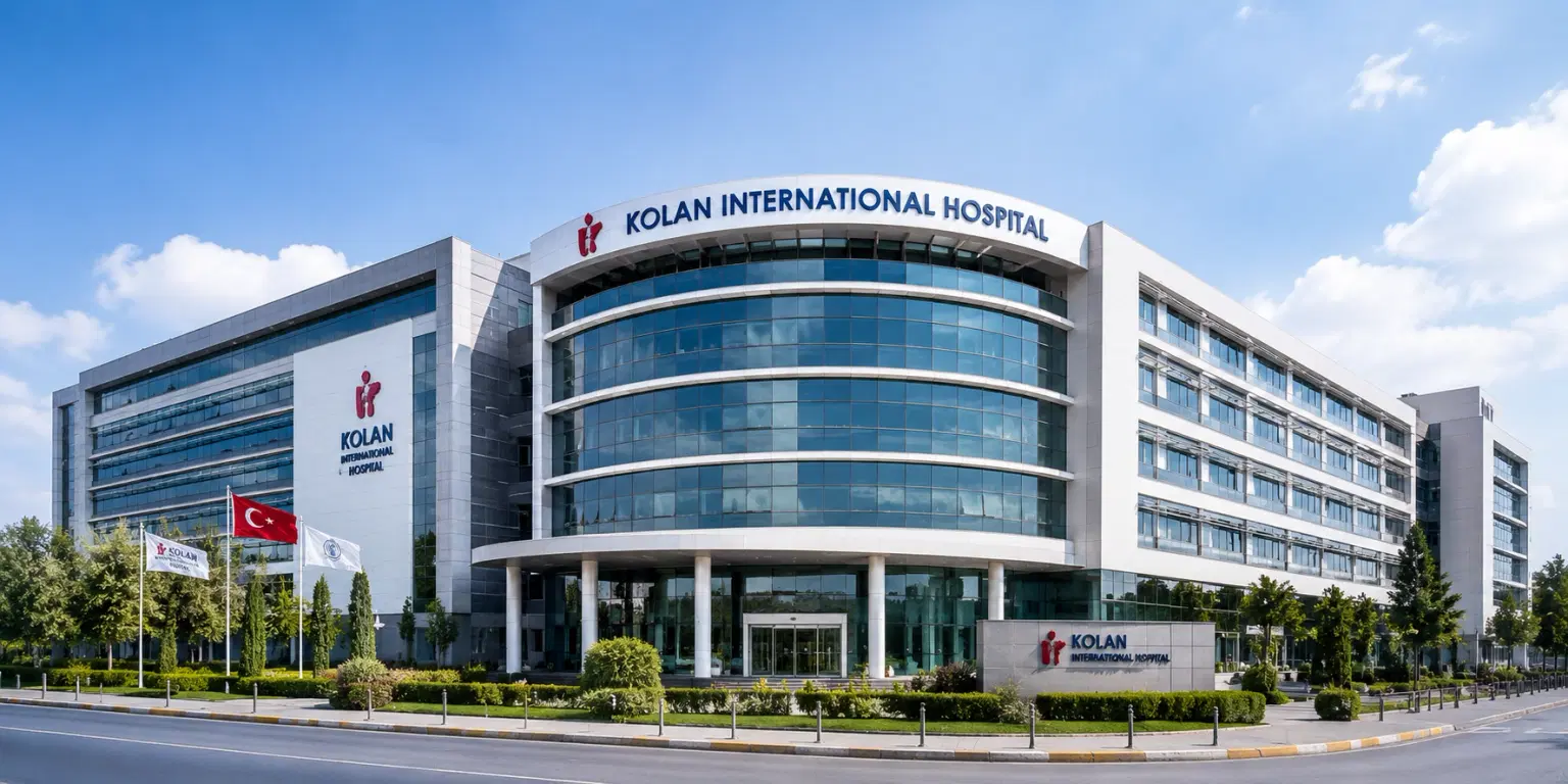
Kolan International Hospital, Istanbul
Kaptanpasa Mah. Okmeydan Kavsagi, Darulaceze Cd. No:14, 34384 Sisli, Istanbul
Kolan International Hospital, Istanbul
Meet Our Doctors
Meet our team of highly qualified and experienced medical professionals dedicated to providing the best healthcare services.
Dr. Abhishek Gupta
Consultant - Internal Medicine
Internal Medicine
New Delhi
12+ Years
Experience
Apolo Delhi
Hospital
1500
Fees

Dr. Arun Puri
Director & Head
Minimal Access, Bariatric & GI Surgery
New Delhi
38+ Years
Experience
Apolo Delhi
Hospital
1500
Fees

Dr. Ashish Vashistha
Principal Consultant
Minimal Access, Bariatric & GI Surgery
New Delhi
23+ Years
Experience
Apolo Delhi
Hospital
1500
Fees

Dr. Atul NC Peters
Chairman, Institute of Bariatric & Metabolic Surgery
Bariatric & Metabolic Surgery
New Delhi
25+ Years
Experience
Apolo Delhi
Hospital
1500
Fees

Dr. Atul Sachdev
Principal Consultant
Gastroenterology
New Delhi
25+ Years
Experience
Apolo Delhi
Hospital
1500
Fees

Dr. Bachan Singh Barthwal
Principal Director - Laparoscopic & Robotic General Surgery
General Surgery, Laparoscopic / Minimal Access Surgery, Robotic Surgery
New Delhi
37+ Years
Experience
Max Hospital,Gurgaon
Hospital
1500
Fees

Dr. Deepa Goel
Head - Histopathology & Cytology Lab
Laboratory Services
New Delhi
21+ Years
Experience
Artemis Hospital
Hospital
1500
Fees

Dr. Devender Yadav
Senior Consultant - Orthopedics (Unit VI)
Orthopaedics
New Delhi
10+ Years
Experience
Artemis Hospital
Hospital
1500
Fees

Dr. Dheeraj Batheja
Senior Consultant - Ortho Spine Surgery
Orthopaedic Surgery, Spine Surgery
New Delhi
12+ Years
Experience
Artemis Hospital
Hospital
1500
Fees

Dr. Ganesh Kumar Mani
Chairman, Cardiac Sciences, Cardiac Surgery (CTVS)
Cardiothoracic and Vascular Surgery (CTVS), Cardiac Sciences
New Delhi
46+ Years
Experience
Max Hospital,Gurgaon
Hospital
1500
Fees
Booking With DIVINHEAL
Get a free consultation to understand your treatment options
Frequently Asked Questions
Get answers to common questions about medical tourism, treatment procedures, and our comprehensive healthcare services.