


heart valve surgery
5K+ International Patients Treated
40+ Source Countries Served
500+ Accredited Partner Hospitals
98% Patient Satisfaction
80% Average Savings vs USA
10K+ Doctors
NABH, JCI Accredited Hospitals
Free Treatment Plan
Free Consultation with Doctor
5+ Destinations Covered
About heart valve surgery
Sources and Guidelines Referenced
The clinical guidance and evidence cited throughout this document originate from major cardiovascular medical societies and landmark peer-reviewed publications: the 2020 AHA/ACC Guideline for the Management of Patients With Valvular Heart Disease (Otto et al., 2021); the 2021 ESC/EACTS Guidelines for the Management of Valvular Heart Disease (Vahanian et al., 2022); NICE Guideline NG208 on Heart Valve Disease in Adults (2021); the PARTNER 3 Trial (Mack et al., New England Journal of Medicine, 2019); the Evolut Low Risk Trial (Popma et al., New England Journal of Medicine, 2019); and the Society of Thoracic Surgeons (STS) Adult Cardiac Surgery Database Reports (2022).
Heart Valve Surgery: A Comprehensive Patient Guide
1. Definition and Medical Identity
Heart valve surgery is a specialized cardiovascular operative procedure designed to correct mechanical dysfunction in one or more of the heart four valves. The primary clinical objective is to restore proper blood flow dynamics, relieve myocardial stress, and improve survival in patients with severe valvular disease.
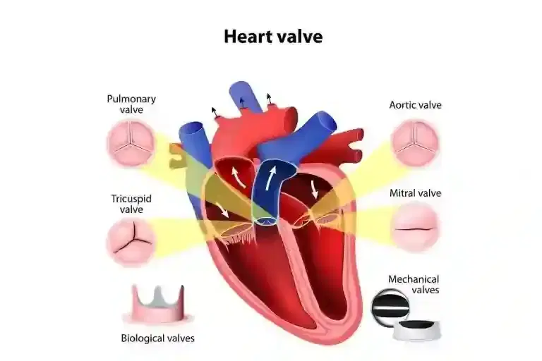
This surgery encompasses two main therapeutic strategies: valve repair, which preserves and reconstructs the patient native tissue, and valve replacement, which substitutes an irreparable native valve with a mechanical or biological prosthesis. Depending on patient risk profiles, anatomical factors, and valve location, operations are performed via traditional median sternotomy (opening the breastbone) or minimally invasive surgical techniques (Vahanian et al., 2022).
2. The Underlying Condition or Need
Heart valve surgery treats structural disease of the cardiac valves that disrupts normal intracardiac hemodynamics. The underlying physiological defect typically presents as either stenosis, where stiffened leaflets restrict forward blood flow, or regurgitation, where incomplete valve closure allows backward blood leakage.
Common clinical presentations include severe dyspnea (shortness of breath), angina (chest pain), syncope (fainting), fatigue, and peripheral edema. Unmanaged valvular dysfunction creates chronic pressure or volume overload on the heart chambers. According to the AHA/ACC guidelines, untreated severe valvular disease progresses to left ventricular dilation, irreversible myocardial dysfunction, secondary pulmonary hypertension, heart failure, and an increased risk of sudden cardiac death (Otto et al., 2021).
3. How the Treatment Works — Mechanism
Heart valve surgery mechanically corrects physical defects that block or divert blood flow. The primary biological aim is to re-establish physiological pressure gradients across cardiac chambers and prevent abnormal backward blood flow.
During surgery, the medical team places the patient on cardiopulmonary bypass. This machine takes over the work of the heart and lungs, diverting unoxygenated venous blood into an external reservoir, oxygenating it, and pumping it back into the arterial system. The surgical team then stops the cardiac cycle using a cold, high-potassium fluid called cardioplegia. This creates a still, bloodless field inside the heart.
For valve repair, the surgeon corrects structural defects using techniques like annuloplasty, where a synthetic ring is sewn around the base of the valve to reduce its size and restore proper leaflet alignment. The surgeon may also trim excess leaflet tissue or replace torn chordae tendineae (fibrous structural cords) with artificial PTFE sutures. For valve replacement, the surgeon cuts away the damaged native leaflets and sews a biological or mechanical prosthetic valve into the structural ring of the original valve.
4. Types and Variations
Surgical protocols vary based on the affected valve, extent of tissue damage, patient age, and bleeding risk. Clinical guidelines emphasize valve repair over replacement whenever technically feasible, particularly for mitral valve disease (Otto et al., 2021).
| Surgical Approach / Type | Primary Mechanism | Key Advantages | Primary Considerations |
|---|---|---|---|
| Valve Repair | Reconstructs native leaflet and chordal structures; places annuloplasty ring. | Preserves native tissue; low blood clot risk; avoids long-term blood thinners. | Technically complex; not feasible for heavily calcified valves. |
| Mechanical Valve Replacement | Implants synthetic valve made from pyrolytic carbon and titanium. | Exceptional durability; lasts 20 to 30+ years without structural failure. | Requires lifelong anticoagulation (warfarin) and daily blood testing. |
| Bioprosthetic (Tissue) Valve | Implants valve constructed from bovine (cow) or porcine (pig) pericardial tissue. | Does not require long-term anticoagulation therapy after initial healing. | Prone to gradual wear; typically lasts 10 to 15 years before replacement. |
| Minimally Invasive Surgery | Accesses heart through minithoracotomy (small rib incision) or upper sternotomy. | Smaller surgical scar; less postoperative pain; faster recovery timeline. | Requires specific vascular access; limited view for multi-valve cases. |
Clinicians select the surgical variation by assessing anatomical suitability on pre-operative imaging, evaluating long-term surgical risks using the Society of Thoracic Surgeons (STS) risk score, and discussing patient preferences regarding blood thinner use and long-term durability (NICE NG208, 2021).
5. Who the Treatment Is For — Indications
Heart valve surgery is indicated for patients with severe, symptomatic structural valve disease, or asymptomatic patients who show signs of declining heart function. Clinical decisions rely on echocardiographic findings, symptom severity, and biomarker trends.
- Severe Symptomatic Valve Stenosis or Regurgitation: Indications include Class II–IV heart failure symptoms (exertional dyspnea, angina, reduced exercise tolerance) directly linked to valve disease (Otto et al., 2021).
- Asymptomatic Left Ventricular Dysfunction: Surgery is indicated for asymptomatic severe mitral or aortic regurgitation when the left ventricular ejection fraction (LVEF) drops below 50–60% or the left ventricle dilates beyond specific thresholds.
- Pulmonary Hypertension: Asymptomatic patients with severe mitral regurgitation who develop resting pulmonary artery systolic pressure greater than 50 mmHg.
- Concomitant Cardiac Surgery: Patients undergoing coronary artery bypass grafting (CABG) or aortic root surgery who also have moderate-to-severe valve disease.
- Infective Endocarditis: Operative intervention is indicated for persistent infection despite antibiotics, large mobile vegetations with high risk of embolism, or severe valve destruction causing acute heart failure (Vahanian et al., 2022).
6. Who the Treatment Is NOT For — Contraindications
Contraindications depend on surgical risk versus expected quality-of-life benefits. Absolute and relative contraindications help clinicians identify patients who may benefit more from medical management or percutaneous interventions.
Absolute contraindications include severe, irreversible non-cardiac comorbidities that limit life expectancy to less than one year, unmanageable acute shock with multi-organ failure, or severe irreversible left ventricular dysfunction where surgery offers no hemodynamic benefit. Relative contraindications include advanced age combined with extreme frailty, calcified "porcelain" aorta (which presents high risks during cross-clamping), severe active systemic infection outside the heart, or severe pulmonary hypertension with right ventricular failure (Vahanian et al., 2022).
7. Alternatives and Clinical Comparison
Medical and percutaneous options exist for patients who are poor candidates for conventional open surgery or who meet specific clinical criteria. Treatment selection depends on multidisciplinary Heart Team evaluations.
| Treatment Option | Mechanism | Invasiveness | Primary Indications | Trade-offs |
|---|---|---|---|---|
| Surgical Valve Repair / Replacement | Direct surgical repair or prosthetic replacement under cardiopulmonary bypass. | High (Open sternotomy or minithoracotomy) | Standard of care for low/intermediate risk patients and complex valve pathology. | Invasive; longer hospital recovery; requires sternal healing period. |
| Transcatheter Aortic Valve Replacement (TAVR) | Deploys a collapsible prosthetic valve via a femoral artery catheter. | Low (Percutaneous vascular catheterization) | Severe aortic stenosis across all surgical risk tiers (Mack et al., 2019). | Higher rate of permanent pacemaker insertion; long-term valve durability still being studied. |
| Transcatheter Edge-to-Edge Repair (TEER / MitraClip) | Clips together excess mitral valve leaflets to reduce backward leak. | Low (Transvenous cardiac catheterization) | Severe symptomatic mitral regurgitation in high surgical risk patients. | Reduces leak but rarely eliminates it completely compared to surgical repair. |
| Medical Management | Pharmacotherapy (diuretics, ACE inhibitors, beta-blockers, anticoagulants). | Non-invasive (Oral medications) | Symptom control, mild/moderate disease, or prohibitive surgical risk. | Controls symptoms but does not repair the physical valve structure. |
8. Pre-Treatment Phase
The pre-treatment phase optimizes patient health and outlines surgical strategy. Initial consultations include detailed medical history taking, functional capacity assessment, and complete physical examination.
The diagnostic workup requires a transthoracic echocardiogram (TTE) supplemented by a transesophageal echocardiogram (TEE) to provide detailed multiplane views of valve anatomy. Cardiac catheterization with coronary angiography evaluates for coexisting coronary artery disease, which may require bypass grafting during the same operation. A chest CT scan assesses aortic calcification and sternal anatomy, while carotid duplex ultrasounds rule out significant carotid artery stenosis that could increase perioperative stroke risk (Otto et al., 2021).
Preoperative preparation also includes a comprehensive dental examination. Any active dental infection or abscess must be treated before elective valve surgery to prevent bacteremia, which can infect the surgical site or prosthetic valve. Medications like antiplatelet agents (e.g., clopidogrel, ticagrelor) and oral anticoagulants are held 3 to 7 days prior to surgery under cardiac oversight. Patients attend pre-admission clinics for blood typing, pulmonary function testing, and instruction on preoperative chlorhexidine skin wash procedures.
9. The Procedure — Step-by-Step Clinical Detail
Heart valve surgery involves a structured sequence of clinical phases executed by a specialized cardiothoracic surgical team.
Phase 1: Induction and Surgical Access
The patient is placed under general anesthesia, and an endotracheal tube is inserted for mechanical ventilation. Invasive monitoring lines are established, including an arterial line for continuous blood pressure monitoring and a central venous line. A transesophageal echocardiography probe is positioned to monitor real-time cardiac performance. The surgeon performs a median sternotomy or a right mini-thoracotomy. The pericardial sac is opened and anchored to expose the heart.
Phase 2: Cardiopulmonary Bypass and Arrest
Heparin is administered intravenously to prevent blood clotting during bypass. Cannulas are placed in the ascending aorta and the right atrium or vena cavae, connecting the patient to the cardiopulmonary bypass machine. Once full mechanical circulatory support is verified, the ascending aorta is cross-clamped to isolate the heart from systemic circulation. Cold cardioplegia solution is infused directly into the coronary arteries, causing immediate, reversible cardiac arrest and preserving myocardial tissue during the operation.
Phase 3: Valve Reconstruction or Replacement
The surgeon makes an incision in the heart or ascending aorta to access the diseased valve. For mitral valve repair, the surgeon may perform leaflet resection, implant artificial PTFE chordae, and secure a rigid or semi-rigid annuloplasty ring along the valve base. For valve replacement, the native valve leaflets and calcified tissue are carefully excised. Sutures are placed around the native valve ring and threaded through the sewing cuff of the mechanical or tissue prosthetic valve. The prosthesis is guided down into position and tied securely.
Phase 4: De-airing, Re-warming, and Decannulation
The cardiac chambers and ascending aorta are filled with blood and vented to purge all air from the system. Re-warming of the body begins. The aortic cross-clamp is removed, restoring blood flow to the coronary arteries. In most cases, the heart spontaneously resumes a normal sinus rhythm; electrical internal defibrillation paddles are used if ventricular fibrillation occurs. Once stable cardiac contractility is confirmed via TEE, cardiopulmonary bypass support is gradually reduced and stopped. Protamine sulfate is administered to reverse the effects of heparin and restore normal blood clotting.
Phase 5: Closure
Chest tubes are inserted into the pericardial and pleural spaces to drain postoperative fluid. Stainless steel wires are threaded around the sternum to close the bone securely. The muscle, subcutaneous tissue, and skin layers are sutured, and sterile dressings are applied.
10. Immediate Post-Procedure Period
The patient transfers directly to the Intensive Care Unit (ICU) under continuous invasive monitoring. Mechanical ventilation continues until the patient awakens, demonstrates adequate muscular strength, meets arterial blood gas criteria, and exhibits minimal chest tube drainage, typically within 4 to 12 hours post-surgery.
Pain management involves continuous intravenous analgesics, patient-controlled analgesia (PCA) pumps, and regional nerve blocks. Clinical staff monitor hourly urine output, chest tube drainage rates, arterial blood pressure, continuous cardiac rhythm, and laboratory values including potassium, hemoglobin, and arterial blood gases. Patients typically transfer from the ICU to a step-down cardiac telemetry floor within 24 to 48 hours once stable and off intravenous vasopressor support.
11. Recovery — Short and Long Term
Recovery spans several stages, transitioning from inpatient mobilization to outpatient cardiac rehabilitation and independent home recovery.
- Days 1 to 2 (ICU to Step-Down Ward): Early bed-side sitting and removal of invasive lines, chest tubes, and urinary catheters as tolerated.
- Days 3 to 7 (Hospital Inpatient): Supervised walking down hallways, progression to independent personal care, transition to oral pain medications, and initiation of oral anticoagulation if indicated. Discharge occurs when pain is controlled, mobility is restored, and cardiac rhythm is stable.
- Weeks 1 to 3 (Early Home Recovery): Daily short walks, breathing exercises using an incentive spirometer, and incision monitoring. Sternal restrictions require patients to avoid pushing, pulling, or lifting items over 10 pounds (4.5 kg).
- Weeks 4 to 8 (Outpatient Rehabilitation): Enrollment in structured cardiac rehabilitation, including supervised cardiovascular exercise, blood pressure monitoring, and nutritional counseling. Patients can typically resume driving around week 6 following clinical sternal check.
- Weeks 9 to 12 and Beyond (Full Recovery): Progressive return to work, non-contact physical activities, and long-term exercise routines. Long-term cardiac imaging evaluates valve function (Otto et al., 2021).
12. Risks, Side Effects, and Complications
Heart valve surgery carries potential early and late clinical complications. Risk levels vary based on underlying comorbidities, patient age, valve position, emergency status, and overall cardiac function.
| Frequency Tier | Side Effect / Complication | Clinical Context and Management |
|---|---|---|
| Common / Mild (10% – 30%) | Postoperative Atrial Fibrillation Surgical Site Discomfort Mild Fluid Retention | Transient cardiac rhythm irregularities treated with beta-blockers or antiarrhythmics; managed with oral analgesics and short-term diuretics. |
| Uncommon (2% – 9%) | Pleural or Pericardial Effusion Acute Kidney Injury (Transient) Wound Healing Complications | Fluid accumulation around lungs or heart; managed conservatively or via catheter drainage. Monitored via serum creatinine. |
| Rare / Serious (< 1% – 2%) | Perioperative Stroke Deep Sternal Wound Infection Complete Heart Block Prosthetic Valve Endocarditis | Neurological deficits from embolic events; deep infection requiring antibiotics and debridement; conduction block requiring permanent pacemaker; infection of implanted valve. |
Surgical mortality rates for isolated elective valve procedures remain low, with STS registry data showing operative mortality rates under 1 to 2% for aortic valve replacement and mitral valve repair at experienced heart centers (STS Database Report, 2022).
Patients must seek urgent medical evaluation if they develop warning signs including fever over 101°F (38.3°C), increased redness or drainage at the surgical incision, sudden shortness of breath, a rapid or irregular pulse, weight gain exceeding 3 pounds in 24 hours, or sudden neurological deficits like weakness or speech difficulty.
13. Lifestyle and Behavioural Considerations
Optimizing operational outcomes requires dedicated lifestyle management before and after surgery. Pre-operative interventions focus on stopping tobacco use at least 4 weeks prior to reduce pulmonary complications, performing recommended physical activity within symptom limits, and optimizing nutritional status.
Postoperative restrictions protect the healing sternum for at least 8 weeks. Patients must avoid pushing up from chairs using arms, lifting heavy weights, or operating motor vehicles until cleared by their surgeon. Long-term management requires strict oral hygiene and prophylaxis guidelines: patients with prosthetic heart valves or repaired valves using prosthetic material must take antibiotic prophylaxis before certain dental procedures to prevent infective endocarditis (Otto et al., 2021).
Patients who receive mechanical valve replacements require lifelong anticoagulation therapy with warfarin to prevent blood clots. This requires maintaining a consistent dietary intake of Vitamin K, along with routine International Normalized Ratio (INR) blood testing to keep anticoagulation within the target therapeutic window (typically 2.0–3.0 for aortic mechanical valves and 2.5–3.5 for mitral mechanical valves).
14. How Outcomes Are Measured
Surgical success is assessed using clinical, functional, and imaging parameters. Short-term endpoints include survival past 30 days, successful weaning from cardiopulmonary bypass, absence of major perioperative neurological events, and complete surgical wound healing.
Long-term functional success is measured using the New York Heart Association (NYHA) functional classification, tracking improvements in symptom severity and daily physical tolerance. Structural success is evaluated via transthoracic echocardiography performed at discharge, 3 months, 12 months, and annually thereafter. Echocardiographic markers assess mean transvalvular pressure gradients, effective valve orifice area, presence and severity of paravalvular regurgitation, and normalization of left ventricular mass and volumes (Vahanian et al., 2022).
Long-term structural valve durability depends on the type of prosthesis implanted. Mechanical valves offer structural durability exceeding 20 to 30 years but carry bleeding risks from required blood thinners. Tissue valves experience progressive structural valve degeneration over time due to calcification and wear, with re-intervention rates increasing 10 to 15 years post-implantation, particularly in younger patients (Otto et al., 2021).
15. Recent Advances and Current Standard of Care
The field of heart valve surgery has advanced considerably over the past two decades. A key trend is the growth of minimally invasive cardiac surgery (MICS) and robotic-assisted valve repair, which allow surgeons to work through smaller incisions between the ribs, reducing surgical trauma and recovery times.
Sutureless and rapid-deployment aortic valves have streamlined biological valve replacement, reducing cardiopulmonary bypass and aortic cross-clamp times. Valve-in-valve transcatheter procedures now allow clinicians to place a new transcatheter valve directly inside a deteriorated tissue valve via percutaneous access, offering a minimally invasive re-intervention option for patients with failing bioprosthetic valves (Popma et al., 2019).
16. Common Myths and Misconceptions
Myth: Heart valve surgery always requires opening the breastbone with a large chest scar.
Reality: Many mitral and aortic valve repairs or replacements are performed using minimally invasive approaches, accessing the heart through small incisions between the ribs without cutting the sternum (Vahanian et al., 2022).
Myth: Tissue valves are always superior to mechanical valves because they do not require blood thinners.
Reality: Bioprosthetic tissue valves degenerate over time and may require re-operation after 10 to 15 years, whereas mechanical valves offer long-term structural durability. The decision involves balancing age, lifestyle factors, and the risks of long-term anticoagulation (Otto et al., 2021).
Myth: Once a heart valve is replaced, the patient is completely cured and requires no further cardiac care.
Reality: Patients require ongoing cardiovascular follow-up, periodic echocardiograms, antibiotic prophylaxis before dental work, and medication management to protect heart function and monitor valve durability.
Myth: Transcatheter approaches like TAVR have completely eliminated the need for open heart valve surgery.
Reality: Surgical valve intervention remains the gold standard for young patients, complex multi-valve pathology, severe mitral repair cases, and individuals with anatomical features unsuitable for percutaneous devices (NICE NG208, 2021).
Myth: You cannot exercise or lead an active life after receiving a heart valve replacement.
Reality: Successful valve repair or replacement restores normal blood flow, allowing most patients to return to active, healthy lifestyles and regular exercise once fully recovered.
Myth: Mechanical valves cause noticeable clicking noises that disrupt daily life.
Reality: While mechanical valves produce a faint closure sound that patients may hear in quiet environments, most individuals adapt quickly, and it does not affect sleep or quality of life.
17. Frequently Asked Questions
What is the difference between heart valve repair and valve replacement?
Heart valve repair preserves and reconstructs your native tissue using techniques like reshaping leaflets or reinforcing the valve base with a ring. Valve replacement removes the damaged valve entirely and substitutes a mechanical or bioprosthetic tissue valve. Guidelines recommend valve repair whenever technically feasible, particularly for mitral valve disease.
How long does a biological tissue valve last compared to a mechanical valve?
Biological tissue valves constructed from animal tissue typically last 10 to 15 years before experiencing wear and calcification. Mechanical valves made from pyrolytic carbon do not wear out structurally and can last 20 to 30 years or longer, though they require lifelong blood-thinning medication.
Why do I need to see a dentist before undergoing heart valve surgery?
Bacteria from active oral infections or dental decay can enter the bloodstream and attach to newly repaired or prosthetic heart valves, causing a severe infection known as endocarditis. Resolving all dental issues prior to surgery significantly reduces this infection risk.
How long will I need to take blood thinners after surgery?
Patients with mechanical valves must take oral anticoagulants like warfarin permanently to prevent blood clots on the valve. Patients with bioprosthetic valves or surgical valve repairs typically take anticoagulants or antiplatelet drugs for 3 to 6 months postoperatively, depending on individual risk factors and heart rhythm.
When can I safely resume driving after heart valve surgery?
Most patients can resume driving approximately 6 weeks after open surgery involving a median sternotomy. This timeline allows the breastbone time to heal structurally, ensuring safe upper body mobility and seatbelt protection. Patients undergoing minimally invasive surgery may return to driving sooner if cleared by their surgeon.
What is sternal precautions and how long do I need to follow them?
Sternal precautions are activity restrictions designed to protect the breastbone while it heals together. Guidelines include avoiding lifting items weighing more than 10 pounds (4.5 kg), avoiding pushing or pulling with your arms, and supporting your chest when coughing or sneezing. These guidelines are usually followed for 6 to 8 weeks post-surgery.
Can a damaged heart valve fix itself or be cured with medication alone?
Structural valve damage cannot heal on its own, and medications cannot un-narrow a stiffened valve or physically repair a leaking leaflet. Medications like diuretics and blood pressure drugs reduce cardiac strain and relieve symptoms, but surgery or percutaneous procedures are necessary to correct mechanical structural defects.
What is an annuloplasty ring and why is it used?
An annuloplasty ring is a specially shaped metal or synthetic ring sewn around the base (annulus) of a damaged heart valve during repair surgery. It restores the proper shape and size of the valve ring, helping the valve leaflets meet correctly to stop backward blood leakage.
How long is the typical hospital stay for heart valve surgery?
The standard hospital stay after heart valve surgery ranges from 5 to 7 days. This includes 1 to 2 days in the intensive care unit for close monitoring, followed by 3 to 5 days on a specialized cardiac step-down ward for physical therapy and medication adjustment.
Will I need antibiotic prophylaxis before dental procedures after valve surgery?
Yes. Clinical guidelines recommend that patients with prosthetic heart valves, bioprosthetic materials, or repairs involving prosthetic rings take single-dose oral antibiotics before invasive dental work. This preventative measure helps protect against bacterial endocarditis.
What are the signs of structural valve degeneration in a bioprosthetic valve?
Structural valve degeneration occurs gradually over years as tissue valve leaflets thicken or calcify. Symptoms mimic original valve disease, including progressive exertional shortness of breath, fatigue, lower limb swelling, or dizziness. Routine annual echocardiograms detect early changes in valve gradients before severe symptoms recur.
Can I travel by airplane after heart valve surgery?
Most patients can travel by air 4 to 6 weeks after surgery, provided recovery is progressing normally and cardiac rhythm is stable. You must consult your cardiovascular team before booking air travel to confirm adequate surgical healing, stable hemoglobin levels, and controlled anticoagulation levels.
What is the STS risk score and how is it used?
The Society of Thoracic Surgeons (STS) risk score is a validated risk assessment tool used by Heart Teams to calculate a patient risk of complications or mortality prior to heart surgery. It evaluates patient age, kidney function, cardiac performance, and comorbidities to guide decisions between open surgery, transcatheter interventions, or medical management.
Booking With DIVINHEAL
Get a free consultation to understand your treatment options
Cost Calculator
I know my treatment - show me cost from 3 hospitals
Plan My Journey
Tell us your condition and budget - our AI matches the right destination, hospital and doctor and visa pathway
Recommended Article

Booking With DIVINHEAL
Get a free consultation to understand your treatment options
Cost Calculator
I know my treatment - show me cost from 3 hospitals
Plan My Journey
Tell us your condition and budget - our AI matches the right destination, hospital and doctor and visa pathway
Recommended Article
Our Speciality and Treatments
oculoplastic surgery
Macular Degeneration Treatment (AMD)
Pediatric Ophthalmology
Squint / Strabismus Surgery (Adult)
ICL / Phakic IOL Implantation
LASIK / SMILE / PRK (Refractive Surgery)
Keratoconus Treatment (CXL / Rings)
Corneal Transplant (Keratoplasty)
Pediatric Squint Surgery
Diabetic Retinopathy Laser / Anti-VEGF Injections
Vitrectomy
Retinal Detachment Repair
Pterygium Surgery
Glaucoma Surgery (Trabeculectomy / MIGS)
Cataract Surgery (Phaco with IOL)






Meet Our Medical Specialists
Hospitals
NABH & JCI Accredited Hospitals in India,Turkey, Thailand & UAE.

Hisar Intercontinental Hospital
Saray Mah. Siteyolu Cad. No:7, Umraniye, 34768, Istanbul, Turkey

Medical Park Group, Istanbul
Fahrettin Kerim Gokay Cad. Tıbbiye Cd., Kadikoy, Istanbul, Turkey

Emsey Hospital, Pendik, Istanbul
Çamlık, Selçuklu Cd. No:22, 34912 Pendik/İstanbul, Türkiye
LIV Hospital, Istanbul
How DivinHeal Helps
We simplify your medical journey by providing comprehensive support and access to world-class healthcare.
Expert Specialist Matching
Connecting you with the world's top-rated medical experts.
Accredited Hospital Network
Access to JCI & NABH certified healthcare facilities.
Complete Travel Coordination
Hassle-free visa, stay, and local transport assistance.
24/7 Personal Care
Dedicated patient advisors supporting you at every step.
Journey Guidance
Full guidance from start to end of the patient treatment journey.
Expert Specialist Matching
Connecting you with the world's top-rated medical experts.
Everything you
need to know today
Browse through these common inquiries to better understand our patient-focused medical platform.
Yes, we work with a variety of insurance providers. Contact our team to verify your coverage.
Yes, we provide secure online consultations with experienced specialists.
Our care coordinators help match you with the most suitable specialist.
Absolutely. Your medical information is protected according to healthcare privacy standards.
Look at six things: accreditation (JCI or NABH), specialty depth, doctor credentials and experience, procedure-specific success rates, international patient support, and technology. DivinHeal's AI-driven matching evaluates every hospital in our accredited partner network on these dimensions and shortlists the best-fit options for your condition, budget, and country.
JCI (Joint Commission International) is the US-based global gold standard for hospital quality, recognised worldwide. NABH is India's national accreditation — accredited by ISQua, the same body that accredits JCI. Both signal independently verified safety and quality. Most of India's leading hospitals hold both.
Yes. All three welcome international patients through structured medical visa programs. India is the most established, treating patients from Africa, the Middle East, and South Asia at 60–80% lower cost. Thailand leads in cosmetic and dental care. The UAE is emerging in oncology and reproductive medicine.
Most patients save 50–80% on treatment costs. Heart bypass costs US $7,000–9,000 in India compared to $70,000–150,000 in the US. IVF costs $3,000–4,500 compared to $12,000–20,000 in the UK. Even after flights, visa, and accommodation, total savings remain 60–70%.
DivinHeal manages your entire non-medical journey: visa invitation letters, medical visa guidance, doctor appointments, teleconsultations, airport pickup, hospital-vetted accommodation for you and your attendant, language interpreters, local transport, cuisine preferences, and post-treatment follow-up — one dedicated coordinator from first enquiry to final follow-up.
You need a valid passport (6+ months validity), a medical visa (M-Visa for India — DivinHeal provides the hospital invitation letter), return flight tickets, recent medical reports and a doctor's referral, current prescription list, and proof of financial means. Any accompanying attendant needs their own passport and MX-Visa.
Still have more questions?
Book a call with our friendly team to learn how DivineHeal simplifies your healthcare journey.