


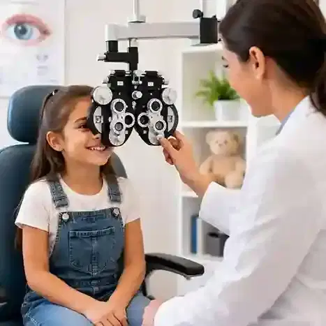
Pediatric Ophthalmology
5K+ International Patients Treated
40+ Source Countries Served
500+ Accredited Partner Hospitals
98% Patient Satisfaction
80% Average Savings vs USA
10K+ Doctors
NABH, JCI Accredited Hospitals
Free Treatment Plan
Free Consultation with Doctor
5+ Destinations Covered
About Pediatric Ophthalmology
Sources and Guidelines Referenced
The clinical claims, developmental thresholds, and protocol summaries in this guide are grounded in established clinical practice guidelines and research published by leading ophthalmic societies: American Association for Pediatric Ophthalmology and Strabismus Guidelines (AAPOS, 2023); American Academy of Ophthalmology Preferred Practice Patterns on Amblyopia (AAO PPP, 2022), Strabismus (AAO PPP, 2020), and Pediatric Eye Evaluations (AAO PPP, 2023); Pediatric Eye Disease Investigator Group multi-center clinical trials (PEDIG, 2018–2023); Royal College of Ophthalmologists Management of Pediatric Cataract Guidelines (RCOphth, 2021); and World Health Organization Guidelines on Prevention of Childhood Blindness (WHO, 2020).
Pediatric Ophthalmology: A Comprehensive Patient Guide
1. Definition and Medical Identity
Pediatric ophthalmology is a subspecialty of medicine focused on visual development, ocular health, and extraocular alignment disorders in infants, children, and adolescents. It combines specialized non-surgical therapies, optical corrections, and subspecialty microsurgical techniques to establish normal visual acuity, preserve binocular vision, and realign eye posture during early visual growth.
The formal medical discipline encompasses the diagnosis and management of conditions affecting the entire visual system, from the cornea and anterior chamber to the retina, optic nerve, and extraocular muscles. Clinicians in this specialty possess training in both general ophthalmology and subspecialty pediatric care, enabling them to evaluate children whose visual systems are actively developing and who cannot articulate visual symptoms.
A core aspect of pediatric ophthalmology is the management of strabismus (misalignment of the eyes) not only in children, but also in adult patients who suffer from congenital or newly developed extraocular muscle imbalances. The goal of intervention is two-fold: restoring optimal anatomical clarity within the visual axis and encouraging normal bilateral neurological processing in the brain's visual cortex.
2. The Underlying Condition or Need
Pediatric eye disorders stem from structural anomalies, uncorrected refractive errors, or extraocular muscle uncoupling during the critical phase of visual neurodevelopment. Without prompt intervention, abnormal visual input prevents the brain from forming clear visual cortical connections, leading to permanent functional visual loss known as amblyopia.
Human visual neurocircuitry is immature at birth. The primary visual cortex requires equal, clear, and simultaneously aligned images from both eyes to develop normal spatial resolution and stereopsis (three-dimensional depth perception). When an eye turn, structural opacity, or unequal refractive power interferes with image quality, the young brain actively suppresses the abnormal image to avoid double vision (diplopia). Over time, this neural suppression leads to structural modifications in the lateral geniculate nucleus and visual cortex, leaving the suppressed eye with reduced visual acuity even if the physical structure of the eye is otherwise healthy.
The natural trajectory of untreated pediatric eye conditions includes permanent unilateral or bilateral visual impairment, loss of depth perception, secondary postural adjustments, and severe functional limitations in later adult life. Conditions such as congenital cataracts or high congenital glaucoma can lead to irreversible blindness if the mechanical barrier to light or elevated pressure is not corrected early in infancy.
3. How the Treatment Works — Mechanism
Pediatric ophthalmology interventions work by clearing the visual axis, focusing sharp light onto the retina, or realigning extraocular muscles. These mechanical and optical adjustments re-establish symmetrical input to the visual cortex, utilizing early neural ocular plasticity to develop normal pathways before critical development windows close.
At the neurological level, treatment relies on the brain's ability to reorganize visual processing centers during early childhood. In amblyopia management, clinicians use mechanical occlusion (eye patching) or pharmacological penalization (atropine drops) on the stronger eye. This forces the visual cortex to process visual signals from the weaker eye, strengthening synaptic connections along the retino-geniculo-striate pathway (Pediatric Eye Disease Investigator Group, 2018).
In strabismus, treatment targets the six extraocular muscles controlling eye movement. Microsurgical techniques alter the physical tension, leverage, or insertion points of these muscles on the sclera (white wall of the eye). Moving a muscle insertion point posteriorly (a recession) relaxes its physical pull, whereas shortening a muscle (a resection or plication) increases its mechanical tension. These alterations adjust the resting eye position, aligning both visual axes to allow binocular fusion in the brain.
4. Types and Variations
Pediatric ophthalmology encompasses non-surgical therapies, diagnostic interventions, and specialized microsurgical procedures. Clinicians select specific protocols based on the child's developmental age, visual acuity, structural anatomy, and underlying condition.
Non-surgical options include optical correction with specialized spectacles, occlusion patching, pharmacologic penalization with cycloplegic drops, and orthoptic visual exercises. Surgical options include strabismus muscle realignment, nasolacrimal duct probing and stenting, pediatric cataract extraction with or without intraocular lens (IOL) placement, goniotomy or trabeculotomy for congenital glaucoma, and laser photocoagulation or anti-VEGF injections for retinopathy of prematurity.
| Category / Procedure | Clinical Mechanism | Target Age Range | Primary Indications | Invasiveness Level |
|---|---|---|---|---|
| Optical Correction | Focuses light sharply on the retina using spectacles or contact lenses | Infants to Adolescents | Anisometropia, high hyperopia, myopia, astigmatism | Non-invasive |
| Occlusion Therapy | Patches the stronger eye to stimulate visual cortex processing from the amblyopic eye | 3 Months to 10 Years | Strabismic, anisometropic, or deprivational amblyopia | Non-invasive |
| Nasolacrimal Probing | Passes a fine metallic wire to clear membranous duct obstruction at the valve of Hasner | 6 Months to 2 Years | Persistent congenital nasolacrimal duct obstruction | Minimally Invasive |
| Strabismus Surgery | Recesses or resects extraocular muscle insertions to balance mechanical eye alignment | 6 Months to Adult | Esotropia, exotropia, vertical strabismus, complex double vision | Surgical (Outpatient) |
| Pediatric Cataract Extraction | Removes opacified crystalline lens, followed by primary posterior capsulotomy and anterior vitrectomy | Birth to Adolescents | Congenital, infantile, or traumatic crystalline lens opacity | Surgical (Inpatient / Outpatient) |
5. Who the Treatment Is For — Indications
Pediatric ophthalmology treatments are indicated for infants, children, and adolescents presenting with eye turns, unequal visual acuity, clear axial obstructions, tear drainage failure, or elevated intraocular pressure. Diagnostic screening guidelines established by the American Academy of Pediatrics (AAP) and AAPOS outline key physical and visual thresholds for specialty referral.
Specific medical indications include:
- Manifest Strabismus: Constant or intermittent eye alignment deviation observed after four months of age (e.g., infantile esotropia or intermittent exotropia).
- Amblyopia Risk Factors: Difference in refractive error between eyes (anisometropia) exceeding 1.50 diopters of hyperopia, 3.00 diopters of myopia, or 1.50 diopters of astigmatism (AAO PPP, 2023).
- Visual Axis Opacities: Any central lens, corneal, or vitreous opacity greater than 3 millimeters in diameter that obstructs light transmission during critical development.
- Congenital Nasolacrimal Duct Obstruction: Epiphora (excessive tearing) and periocular crusting persisting past 12 months of age despite conservative massage.
- Retinopathy of Prematurity: Premature infants born at or under 30 weeks gestation or with birth weight under 1,500 grams requiring retinal vascular evaluation.
6. Who the Treatment Is NOT For — Contraindications
Absolute and relative contraindications in pediatric ophthalmology depend on the surgical or therapeutic approach. Interventions must balance visual benefits against systemic risks associated with pediatric anesthesia and physical compliance capacity.
Absolute contraindications for elective extraocular muscle or optical surgery include severe, non-correctable structural visual pathway absence (such as complete optic nerve aplasia or bilateral anophthalmos) where no visual potential exists. Surgical intervention is also contraindicated in patients with acute, untreated intraocular infections (endophthalmitis) or systemic medical instability that makes general anesthesia life-threatening.
Relative contraindications include poor patient or caregiver ability to comply with post-operative topical medication schedules or eye patching regimes. Uncontrolled active ocular surface inflammation, severe dry eye syndrome, or unstable progressive systemic metabolic disorders require medical stabilization prior to elective eye procedures.
7. Alternatives and Clinical Comparison
Alternative approaches exist across most pediatric ophthalmology management pathways. Clinicians evaluate trade-offs between invasiveness, daily lifestyle burden, compliance requirements, and treatment speed when recommending a plan.
For amblyopia, atropine penalization therapy serves as the major clinical alternative to mechanical patching. Randomized multi-center trials by the Pediatric Eye Disease Investigator Group (PEDIG) demonstrate that daily or weekend administration of 1% atropine eye drops in the non-amblyopic eye achieves visual acuity gains equivalent to 2 to 6 hours of daily occlusion patching in mild to moderate amblyopia (PEDIG, 2018).
| Clinical Indication | Primary Intervention | Alternative Approach | Efficacy Comparison | Compliance Requirements | Key Trade-Offs |
|---|---|---|---|---|---|
| Amblyopia | Occlusion (Patching) Therapy | Pharmacologic (Atropine) Penalization | Equivalent visual acuity improvement in mild/moderate cases | Patching requires active physical compliance; Atropine requires eye drop drop administration | Patching causes skin irritation; Atropine causes light sensitivity and blurred near vision in good eye |
| Accommodative Esotropia | Hyperopic Spectacle Prescription | Strabismus Muscle Surgery | Spectacles eliminate turn without surgery when high hyperopia is fully corrected | Daily spectacle wear mandatory | Spectacles avoid surgical risk; surgery without spectacles can cause secondary consecutive exotropia |
| Nasolacrimal Duct Obstruction | Surgical Duct Probing & Irrigation | Conservative Sac Massage (Crigler technique) | Probing has >90% success after age 1; Massage resolves ~80% before age 1 | Massage requires daily parental application for months | Massage is non-invasive; probing requires brief pediatric anesthesia |
8. Pre-Treatment Phase
The pre-treatment phase focuses on comprehensive clinical evaluation, diagnostic imaging, refractive testing, and family preparation. The examination is adapted to the child's developmental age using specialized, non-verbal assessment tools.
Visual acuity in non-verbal infants is assessed using preferential looking techniques, such as Teller Acuity Cards or LEA Symbols. Visual alignment is evaluated using light reflex testing (Hirschberg and Krimsky tests) and cover/uncover testing. Diagnostic evaluation requires cycloplegic refraction. Clinicians instill drops such as cyclopentolate 1% or atropine to temporarily paralyze the ciliary muscle (blocking accommodation) and dilate the pupil. This allows accurate measurement of underlying refractive errors without interference from the child's natural focusing effort (AAO PPP, 2023).
When surgery is planned, pre-operative protocols include a pediatric general health assessment, blood panels if indicated by underlying systemic disease, and coordination with a pediatric anesthesiologist. Fasting (NPO) guidelines must be strictly followed to prevent aspiration during anesthesia. Caregivers receive targeted education regarding post-operative drops, eye protection shields, and activity limitations.
9. The Procedure — Step-by-Step Clinical Detail
Pediatric ophthalmic surgery takes place in a dedicated surgical suite using an operating microscope. Pediatric patients undergo general anesthesia administered by a specialist anesthesiologist using endotracheal intubation or a laryngeal mask airway.
Step 1: Preparation and Exposure
The child is positioned supine, and povidone-iodine antiseptic solution is applied to the ocular surface and periocular skin. A sterile surgical drape isolates the eye, and a specialized pediatric wire speculum is inserted to gently separate the eyelids.
Step 2: Conjunctival Incision and Muscle Identification (Strabismus Surgery)
The surgeon makes a microscopic incision through the conjunctival tissue near the limbus or in the fornix (limbal or fornix approach). Muscle calipers isolate the targeted extraocular muscle (such as the medial rectus). Muscle sutures are passed through the muscle insertion site using fine absorbable thread (e.g., 6-0 or 7-0 polyglactin 910).
Step 3: Muscle Recession or Resection
For a muscle recession (weakening procedure), the muscle is detached from the sclera, measured backward a precise number of millimeters using specialized calipers, and re-anchored to the sclera at its new position. For a muscle resection (strengthening procedure), a segment of the muscle is measured, clamped, excised, and the shortened muscle is reattached to its original insertion site.
Step 4: Nasolacrimal Duct Probing (If Indicated)
For tear duct obstruction, the surgeon dilates the lacrimal punctum with a fine dilator. A smooth Bowman metallic probe is inserted down through the canaliculus, turned vertical, and advanced through the nasolacrimal duct into the inferior meatus of the nose, popping through the obstructed membrane at the valve of Hasner. Fluorescein-dyed saline is irrigated through the duct to confirm clearance under direct visualization.
Step 5: Pediatric Cataract Extraction (If Indicated)
For pediatric cataracts, clear corneal micro-incisions are created. Phacoemulsification or aspiration removes the opacified crystalline lens. Crucially, in pediatric eyes, a primary posterior capsulotomy and anterior vitrectomy are performed to prevent rapid secondary posterior capsule opacification (RCOphth, 2021). An intraocular lens (IOL) is implanted if the child is over 1 to 2 years of age; infants younger than one year are frequently left aphakic and fitted with post-operative contact lenses.
Step 6: Closure and Postoperative Shielding
Conjunctival incisions are closed with delicate absorbable sutures or allowed to self-seal. Antibiotic and corticosteroid ointments are applied, and a protective rigid plastic shield is secured over the eye to prevent accidental touch during recovery.
10. Immediate Post-Procedure Period
The immediate post-procedure period encompasses the first 24 to 48 hours following surgery. Patients are monitored in the Post-Anesthesia Care Unit (PACU) until general anesthesia resolves and systemic vital signs stabilize.
Normal immediate post-operative findings after extraocular muscle surgery include moderate conjunctival injection (redness), mild periorbital edema, transient pink-tinged tearing, and mild scratchy discomfort. Ocular pain in children is typically mild and effectively managed with over-the-counter pediatric paracetamol (acetaminophen) or ibuprofen.
Discharge criteria require the patient to be alert, displaying stable vital signs, tolerating oral fluids without nausea, and showing clear wound site stability without active bleeding. Caregivers receive specific instructions on instilling topical anti-inflammatory and antibiotic drops three to four times daily and applying a protective eye shield during sleep.
11. Recovery — Short and Long Term
Recovery spans short-term physical healing of ocular tissues and long-term visual cortex rehabilitation. Complete functional recovery relies on consistent post-operative topical drop routines and follow-up visual acuity re-assessments.
Short-Term Timeline (Days 1 to 14)
During the first week, redness and local conjunctival swelling gradually decrease. Eye drops containing broad-spectrum antibiotics and steroids (e.g., tobramycin/dexamethasone) are administered for 7 to 14 days. Children may return to school or daycare within 3 to 7 days once comfortable. Active swimming, rough contact play, sandboxes, and eye rubbing must be strictly avoided for at least two weeks to reduce infection risks.
Medium-Term Timeline (Weeks 2 to 8)
Absorbable sutures dissolve completely over 2 to 6 weeks, during which minor localized itching may occur. Final anatomical alignment of extraocular muscles stabilizes by week six. An initial post-operative alignment check is conducted between 2 and 6 weeks to evaluate surgical success and determine if residual double vision or misalignment remains.
Long-Term Timeline (Months 3 to Years)
Long-term care involves monitoring visual acuity development, binocular vision fusion, and refractive state updates every 3 to 6 months until visual maturity (age 8 to 10). If amblyopia persists post-alignment, patching regimens are re-initiated to optimize spatial visual acuity gains.
12. Risks, Side Effects, and Complications
Although pediatric ophthalmic interventions are safe, procedures carry recognized surgical, optical, and anesthetic risks. The risk severity matrix below details complications, frequencies, and management strategies based on published clinical data.
| Severity Tier | Potential Complication | Estimated Frequency | Clinical Presentation | Management Strategy |
|---|---|---|---|---|
| Common / Mild | Conjunctival redness & swelling | >90% post-surgery | Localized redness, watery tearing, mild grit sensation | Self-limiting over 10–14 days; managed with lubricating and steroid drops |
| Common / Mild | Skin irritation from patching | 15% – 25% | Erythema on cheek/periocular skin during occlusion therapy | Switching to hypoallergenic adhesives or silicone-based patches |
| Uncommon | Alignment Over- or Under-correction | 10% – 20% | Residual eye turn (esotropia/exotropia) 6 weeks post-surgery | Prism spectacles, optical correction, or secondary surgical adjustment |
| Uncommon | Granuloma or Scleral Dellen | 1% – 3% | Focal elevated pink tissue on conjunctiva or localized corneal thinning | Topical steroid therapy or minor surgical excision of granuloma |
| Rare / Serious | Endophthalmitis (Internal Infection) | < 0.05% | Severe intraocular pain, sudden vision drop, hypopyon, deep inflammation | Emergency intravitreal antibiotic injections and potential vitrectomy |
| Rare / Serious | Scleral Perforation / Retinal Detachment | < 0.1% | Inadvertent pass of needle into vitreous cavity during muscle reattachment | Intraoperative indirect ophthalmoscopy, cryotherapy, or retinal repair |
Severe complications such as intraocular infection (endophthalmitis) or retinal detachment are rare (<0.1%) when standardized sterile operating room techniques are maintained. Anesthesia complications in pediatric patients are minimized by using specialized pediatric anesthesiology teams and monitoring protocols (AAPOS Guidelines, 2023).
13. Lifestyle and Behavioural Considerations
Successful pediatric eye outcomes rely heavily on behavioral adaptation and daily compliance with optical or pharmacological therapies. Because young children rarely recognize their own visual deficits, parents play a central role in managing therapy routines.
For children undergoing occlusion therapy, integrating patching schedules into structured daily activities—such as reading, crafts, or supervised screen time—improves adherence. Using incentive reward charts and engaging teachers to support patching during school hours increases compliance rates (PEDIG, 2018).
Children who wear high-prescription spectacles or have undergone ocular surgery should wear protective poly-carbonate lenses during sports and physical play to shield against impact injuries. Digital screen usage should follow standard pediatric recommendations: balanced near-work breaks (the 20-20-20 rule: taking a 20-second break every 20 minutes) to prevent digital eye strain and accommodating fatigue, alongside outdoor natural light exposure to reduce myopia progression rates.
14. How Outcomes Are Measured
Clinical outcomes in pediatric ophthalmology are measured using objective visual acuity benchmarks, orthoptic motor alignment tests, and binocular sensory testing. Long-term success is defined as functional, stable vision and aligned binocular integration.
Visual acuity is quantified using standard logarithmic charts, such as LogMAR, LEA Symbols, or Snellen charts, adjusted for the child's age. Symmetrical spatial acuity (within one LogMAR line difference between eyes) signifies successful amblyopia treatment. Motor alignment is measured in prism diopters using alternate prism cover testing at distance and near fixation targets. Surgical success in strabismus is clinically defined as resting ocular alignment within 8 to 10 prism diopters of orthophoria (straight alignment).
Sensory outcomes are evaluated using stereoscopic vision tests, such as the Titmus Fly or Randot Stereo Test. Achieving measurable stereopsis confirms that the visual cortex is processing overlapping input from both eyes simultaneously. In cases of complex childhood strabismus or congenital cataracts, long-term outcome models demonstrate that 15% to 25% of patients may require secondary re-operations or ongoing optical adjustments as their facial and orbital anatomy grows.
15. Recent Advances and Current Standard of Care
Pediatric ophthalmology has advanced significantly through binocular digital treatment technologies, precise intraocular lens manufacturing, advanced retinal imaging, and evidence-based clinical protocols established by multi-center research networks.
A major development in amblyopia therapy is the introduction of binocular dichoptic digital games and virtual reality software. These systems project high-contrast images to the amblyopic eye while projecting lower-contrast images to the non-amblyopic eye, encouraging both eyes to work together binocularly rather than relying on mono-ocular occlusion alone (PEDIG, 2021). Clinical trials demonstrate that dichoptic treatment offers a viable adjunct or alternative to traditional patching in select pediatric populations.
In the management of retinopathy of prematurity (ROP), intravitreal anti-VEGF (vascular endothelial growth factor) agents—such as ranibizumab or bevacizumab—have expanded treatment options alongside conventional laser photocoagulation (RAINBOW Trial, 2019). Anti-VEGF therapy allows continued normal peripheral retinal vascularization in high-risk infants, reducing rates of permanent peripheral visual field loss associated with extensive destructive laser treatments.
16. Common Myths and Misconceptions
Myth: Children will automatically outgrow crossed or misaligned eyes over time.
Reality: True strabismus present past four months of age does not resolve naturally. Delaying clinical evaluation increases the risk of permanent amblyopia and loss of depth perception (AAO PPP, 2020).
Myth: Infants and toddlers are too young to have a comprehensive eye exam or wear glasses.
Reality: Pediatric ophthalmologists can perform complete diagnostic visual evaluations and cycloplegic refractions on newborn infants using objective optical instruments, prescribing specialized infant frames when indicated.
Myth: Eye patching cures lazy eye permanently within a few days of treatment.
Reality: Amblyopia treatment requires consistent therapy over months or years. Rapidly stopping treatment can cause visual acuity regression (PEDIG, 2018).
Myth: Vision screening at a pediatrician's office or school is sufficient to replace a specialist eye examination.
Reality: Basic vision screenings detect gross visual changes but miss high hyperopia, subtle strabismus, or early retinal conditions that require cycloplegic evaluation by an eye specialist.
Myth: Strabismus surgery is purely cosmetic and offers no functional benefits to the child.
Reality: Realigning the eyes restores binocular fusion, establishes stereopsis (3D vision), expands peripheral visual fields, and supports normal psychosocial development.
Myth: Eye exercises alone can eliminate high refractive errors or replace eye surgery for structural muscle imbalances.
Reality: Orthoptic visual therapy can improve convergence insufficiency, but it cannot change the shape of the eyeball (refractive error) or fix structural extraocular muscle misalignments requiring surgery.
Myth: Eating large amounts of carrots will correct childhood vision problems and eliminate the need for glasses.
Reality: While vitamin A supports normal retinal function, dietary intake cannot alter structural shape errors like myopia, hyperopia, or astigmatism.
17. Frequently Asked Questions
At what age should a child have their first comprehensive eye examination?
Infants receive basic red reflex and ocular inspection at birth and during routine pediatric checkups. A comprehensive examination by a pediatric ophthalmologist is recommended if a child fails a vision screening, exhibits an eye turn after four months, or has high-risk conditions such as prematurity, family history of childhood eye disease, or developmental delays (AAO PPP, 2023).
How can an ophthalmologist examine a baby who cannot talk or read letters?
Pediatric ophthalmologists use objective, non-verbal diagnostic tools. Visual acuity is assessed using preferential looking cards (Teller Acuity Cards) that monitor an infant's natural reflex to look at structured patterns. Eye alignment is tested using light reflections and cover tests, while refractive errors are measured using retinoscopy after cycloplegic eye drops temporarily paralyze the focusing muscles.
What is the difference between an optometrist, an ophthalmologist, and a pediatric ophthalmologist?
An optometrist provides primary vision care, optical prescriptions, and general vision testing. An ophthalmologist is a medical doctor (MD or DO) who performs medical and surgical eye care. A pediatric ophthalmologist is a medical doctor who completed additional fellowship training specifically focused on children's visual development, extraocular muscle disorders, and pediatric eye surgery.
How many hours a day does a child need to wear an eye patch for amblyopia?
Prescribed patching duration depends on the severity of amblyopia. Clinical trials by the Pediatric Eye Disease Investigator Group (PEDIG) demonstrate that two hours of daily active patching is as effective as six hours for moderate amblyopia. Severe amblyopia typically requires six hours of daily patching. Clinical follow-up guides individual patching schedules.
Is strabismus surgery safe for infants and young children?
Strabismus surgery is a safe, routinely performed outpatient procedure. Modern pediatric anesthesia, specialized microsurgical instruments, and precise extraocular muscle techniques minimize systemic and ocular risks. While general surgical and anesthetic risks exist, serious complications occur in less than 1% of cases (AAPOS Guidelines, 2023).
Will my child need to wear glasses permanently after strabismus surgery?
Strabismus surgery adjusts the extraocular muscles to realign the eyes; it does not change the physical shape of the cornea or eyeball. If your child had refractive errors such as farsightedness, nearsightedness, or astigmatism prior to surgery, they will still need to wear spectacles after operation to maintain clear visual focus.
How quickly does a child recover after surgery for crossed eyes?
Initial recovery occurs within 24 to 48 hours, during which children experience mild ocular discomfort and eye redness. Most children resume light normal activities and return to school within 3 to 7 days. Complete surface healing and final eye muscle alignment settle over 4 to 6 weeks following surgery.
What happens if amblyopia is not treated before the age of eight or nine?
Visual cortical plasticity decreases as the brain matures around age eight to ten. Delaying amblyopia treatment beyond this critical development window makes visual restoration significantly more challenging, often leading to permanent visual acuity loss in the affected eye that cannot be corrected in adulthood (AAO PPP, 2022).
Can atropine drops effectively replace eye patching for lazy eye?
Yes. Clinical trials confirm that daily or weekend administration of 1% atropine drops in the stronger eye provides visual acuity improvements equivalent to occlusion patching in children with mild to moderate amblyopia. Atropine temporarily blurs the near vision of the strong eye, forcing the brain to use the amblyopic eye.
How do clinicians unblock a clogged tear duct in an infant?
Initial management involves conservative Crigler tear sac massage and warm compresses during the first year of life, which resolves approximately 80% of congenital obstructions. If tearing persists past 12 months, a brief outpatient surgical probing is performed under light anesthesia to clear the thin membrane obstructing the duct.
What are the warning signs that a child may have a serious visual problem?
Warning signs include persistent inward or outward eye turning after four months of age, a white reflex inside the pupil on flash photos (leukocoria), excessive tearing, extreme sensitivity to light, constant eye rubbing, head tilting while focusing, or squinting when viewing distant objects.
Why are dilating drops necessary during a pediatric eye examination?
Dilating drops temporarily relax the eye's internal ciliary muscle (blocking accommodation) and enlarge the pupil. Children have powerful focusing muscles that mask underlying farsightedness or astigmatism. Cycloplegia allows clinicians to obtain an accurate objective measurement of refractive error and perform a thorough examination of the internal retina and optic nerve.
Can pediatric cataracts occur in newborn babies?
Yes. Congenital cataracts can be present at birth due to genetic mutations, metabolic disorders, intra-uterine infections (such as rubella or cytomegalovirus), or structural ocular malformations. Prompt surgical extraction within the first 6 to 8 weeks of life is critical to prevent severe deprivational amblyopia.
Will playing video games or looking at screens damage a child's eyes?
Digital screen use does not cause permanent organic damage to extraocular structures, but excessive close screen work causes digital eye strain, temporary focusing fatigue, and dry eye symptoms. Increased time spent on close near-work combined with reduced outdoor exposure is associated with higher rates of myopia progression in growing children.
Booking With DIVINHEAL
Get a free consultation to understand your treatment options
Cost Calculator
I know my treatment - show me cost from 3 hospitals
Plan My Journey
Tell us your condition and budget - our AI matches the right destination, hospital and doctor and visa pathway
Recommended Article

Booking With DIVINHEAL
Get a free consultation to understand your treatment options
Cost Calculator
I know my treatment - show me cost from 3 hospitals
Plan My Journey
Tell us your condition and budget - our AI matches the right destination, hospital and doctor and visa pathway
Recommended Article
Our Speciality and Treatments
oculoplastic surgery
Macular Degeneration Treatment (AMD)
Pediatric Ophthalmology
Squint / Strabismus Surgery (Adult)
ICL / Phakic IOL Implantation
LASIK / SMILE / PRK (Refractive Surgery)
Keratoconus Treatment (CXL / Rings)
Corneal Transplant (Keratoplasty)
Pediatric Squint Surgery
Diabetic Retinopathy Laser / Anti-VEGF Injections
Vitrectomy
Retinal Detachment Repair
Pterygium Surgery
Glaucoma Surgery (Trabeculectomy / MIGS)
Cataract Surgery (Phaco with IOL)






Meet Our Medical Specialists
Hospitals
NABH & JCI Accredited Hospitals in India,Turkey, Thailand & UAE.
How DivinHeal Helps
We simplify your medical journey by providing comprehensive support and access to world-class healthcare.
Expert Specialist Matching
Connecting you with the world's top-rated medical experts.
Accredited Hospital Network
Access to JCI & NABH certified healthcare facilities.
Complete Travel Coordination
Hassle-free visa, stay, and local transport assistance.
24/7 Personal Care
Dedicated patient advisors supporting you at every step.
Journey Guidance
Full guidance from start to end of the patient treatment journey.
Expert Specialist Matching
Connecting you with the world's top-rated medical experts.
Everything you
need to know today
Browse through these common inquiries to better understand our patient-focused medical platform.
Yes, we work with a variety of insurance providers. Contact our team to verify your coverage.
Yes, we provide secure online consultations with experienced specialists.
Our care coordinators help match you with the most suitable specialist.
Absolutely. Your medical information is protected according to healthcare privacy standards.
Look at six things: accreditation (JCI or NABH), specialty depth, doctor credentials and experience, procedure-specific success rates, international patient support, and technology. DivinHeal's AI-driven matching evaluates every hospital in our accredited partner network on these dimensions and shortlists the best-fit options for your condition, budget, and country.
JCI (Joint Commission International) is the US-based global gold standard for hospital quality, recognised worldwide. NABH is India's national accreditation — accredited by ISQua, the same body that accredits JCI. Both signal independently verified safety and quality. Most of India's leading hospitals hold both.
Yes. All three welcome international patients through structured medical visa programs. India is the most established, treating patients from Africa, the Middle East, and South Asia at 60–80% lower cost. Thailand leads in cosmetic and dental care. The UAE is emerging in oncology and reproductive medicine.
Most patients save 50–80% on treatment costs. Heart bypass costs US $7,000–9,000 in India compared to $70,000–150,000 in the US. IVF costs $3,000–4,500 compared to $12,000–20,000 in the UK. Even after flights, visa, and accommodation, total savings remain 60–70%.
DivinHeal manages your entire non-medical journey: visa invitation letters, medical visa guidance, doctor appointments, teleconsultations, airport pickup, hospital-vetted accommodation for you and your attendant, language interpreters, local transport, cuisine preferences, and post-treatment follow-up — one dedicated coordinator from first enquiry to final follow-up.
You need a valid passport (6+ months validity), a medical visa (M-Visa for India — DivinHeal provides the hospital invitation letter), return flight tickets, recent medical reports and a doctor's referral, current prescription list, and proof of financial means. Any accompanying attendant needs their own passport and MX-Visa.
Still have more questions?
Book a call with our friendly team to learn how DivineHeal simplifies your healthcare journey.