


Keratoconus Treatment (CXL / Rings)
5K+ International Patients Treated
40+ Source Countries Served
500+ Accredited Partner Hospitals
98% Patient Satisfaction
80% Average Savings vs USA
10K+ Doctors
NABH, JCI Accredited Hospitals
Free Treatment Plan
Free Consultation with Doctor
5+ Destinations Covered
About Keratoconus Treatment (CXL / Rings)
Sources and Guidelines Referenced
Every clinical statement, protocol requirement, and safety threshold in this document is grounded in established ophthalmic literature and professional consensus guidelines, including the American Academy of Ophthalmology (AAO) Preferred Practice Pattern for Keratoconus and Corneal Ectasia (2023), the Global Consensus on Keratoconus and Ectatic Diseases (Gomes et al., 2015), the National Institute for Health and Care Excellence (NICE) Interventional Procedures Guidance [IPG404], and foundational clinical trial publications (Wollensak et al., 2003; Raiskup et al., 2015; Hashemi et al., 2020).
Keratoconus Treatment (CXL / Rings): A Comprehensive Patient Guide
1. Definition and Medical Identity
Corneal collagen cross-linking (CXL) and intrastromal corneal ring segments (ICRS) are targeted ophthalmic procedures used to stabilize and structurally modify a diseased cornea (the clear front window of the eye). CXL is a photochemical procedure designed to halt ectatic progression, while ICRS placement is a structural micro-surgical procedure designed to reshape the irregular corneal curvature.
Together, these therapeutic modalities fall under the specialty of cornea and external disease within ophthalmology. CXL is referred to clinically as photochemical corneal collagen cross-linking, transepithelial cross-linking, or accelerated cross-linking. Intrastromal ring segments are commonly known by their trade or technical designations, including Intacs, Keraring, or Ferrara rings. Unlike corrective lenses, which merely compensate for refractive errors, these interventions directly address the structural and biomechanical integrity of the corneal tissue.
2. The Underlying Condition or Need
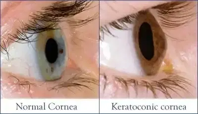
Keratoconus is a non-inflammatory, progressive structural condition characterized by localized thinning and outward structural weakening of the corneal stroma (the thick middle structural layer of the eye). Intraocular pressure pushes this weakened tissue outward, altering the normal spherical curve of the eye into an asymmetric, cone-like protrusion.
This progressive shape change disrupts how light focuses onto the retina, causing irregular astigmatism (uneven curvature of the optical surface), progressive myopia (nearsightedness), higher-order optical aberrations, optical glare, and distorted double vision. If left untreated, progressive keratoconus leads to persistent loss of best-corrected visual acuity, corneal scarring, structural hydrops (fluid leakage into the stroma following tissue rupture), and eventual reliance on high-risk full-thickness corneal transplantation.
3. How the Treatment Works — Mechanism
CXL and ICRS stabilize and reshape the eye through separate photodynamic and biomechanical mechanisms that reinforce weak corneal tissues.
The biological mechanism of CXL relies on photodynamic excitation. When riboflavin (vitamin B2) saturates the corneal stroma and is exposed to controlled 365 nm ultraviolet-A light, it absorbs photon energy and generates reactive oxygen species. These free radicals induce photo-oxidation reactions, creating new covalent chemical bonds between collagen amino acid residues and surrounding proteoglycans. This biochemical cross-linking stiffens the corneal matrix, stopping progressive bulging (Wollensak et al., 2003).
The mechanism of ICRS relies on mechanical structural displacement. Inserting rigid PMMA arc-segments into deep peripheral stromal channels alters local tension. According to Barraquer's tissue addition law, adding mass to the corneal periphery flattens the central tissue proportional to the thickness of the inserted segment. This flattens the apex of the cone, improves optical symmetry, and reduces high-degree irregular astigmatism.
4. Types and Variations
Clinical protocols for both CXL and ICRS are tailored to patient age, disease severity, corneal thickness, and corneal mapping patterns.
CXL protocols are broadly categorized by how the surface skin of the eye—the epithelium—is managed, and by the intensity of UVA light applied. The conventional Dresden protocol involves removing the central epithelium (Epi-off) to ensure deep riboflavin absorption, followed by 30 minutes of UVA exposure at 3 mW/cm². Accelerated Epi-off CXL delivers higher intensity energy over a shorter duration (such as 9 mW/cm² for 10 minutes), yielding comparable biomechanical stabilization (Hashemi et al., 2020). Transepithelial (Epi-on) CXL leaves the surface layer intact using chemical enhancers, reducing postoperative pain but demonstrating lower cross-linking density in clinical studies.
ICRS variations are classified by segment thickness, arc length, and structural cross-section. Surgeons select specific arc lengths (ranging from 90 to 210 degrees) and variable thicknesses (150 to 350 micrometers) depending on whether the astigmatism is symmetric or asymmetric.
| Treatment Variation | Epithelial Handling / Channel Creation | Primary Mechanism | Clinical Indications | Key Trade-Offs |
|---|---|---|---|---|
| Standard Epi-Off CXL (Dresden) | Manual epithelial removal (8–9 mm) | Photochemical covalent bond formation via 3 mW/cm² UVA for 30 min | Documented progressive keratoconus; pachymetry >400 µm | Highest evidence base; requires 3–5 day epithelial healing phase with temporary pain |
| Accelerated Epi-Off CXL | Manual epithelial removal (8–9 mm) | Photochemical bond formation via 9–30 mW/cm² UVA for 3–10 min | Progressive keratoconus in patients needing shorter procedure times | Faster treatment duration; equivalent long-term stabilization outcomes |
| Epi-On (Transepithelial) CXL | Epithelium kept intact; enhanced formula | Deeper chemical penetration without surface debridement | Mild progression; low pain tolerance; pediatric patients | Significantly less post-op discomfort; lower total cross-linking depth |
| ICRS (Intrastromal Rings) | Femtosecond laser or manual stromal channels | Mechanical flattening of central cone via tissue addition | Moderate keratoconus with irregular astigmatism and lens intolerance | Improves uncorrected vision; carries low risk of channel infection or extrusion |
5. Who the Treatment Is For — Indications
Candidates for CXL or ICRS undergo rigorous clinical evaluation using high-definition anterior segment imaging. Treatment selection depends on documented disease progression and functional visual limits.
- Documented Disease Progression: An increase in maximum corneal curvature (Kmax) of 1.0 Diopter or more over 12 months, progressive stromal thinning, or documented refractive shifts (AAO PPP, 2023).
- Pediatric Keratoconus: Children and adolescents under 18 diagnosed with keratoconus often skip the 12-month monitoring period and undergo prompt CXL, as youthful tissue tends to progress rapidly.
- Contact Lens Intolerance: Patients whose severe corneal asymmetry prevents stable fitting of rigid gas permeable or scleral contact lenses are prime candidates for flattening with ICRS.
- Post-Refractive Surgery Ectasia: Patients experiencing progressive structural weakening following previous LASIK or PRK procedures.
- Adequate Stromal Thickness: For safe CXL delivery without injuring the delicate inner endothelium (the pump cells lining the back of the cornea), stromal thickness must measure at least 400 micrometers after epithelial removal.
6. Who the Treatment Is NOT For — Contraindications
Certain ocular and systemic medical conditions prevent safe execution of CXL or ICRS, requiring alternative clinical approaches.
- Corneal Thickness Below Threshold: A thin stromal bed measuring less than 400 micrometers (even after hypo-osmolar swelling protocols) is an absolute contraindication for standard CXL, as UVA radiation risks permanently damaging the inner endothelial cell layer.
- Advanced Central Corneal Scarring: Dense, permanent central optical scars cannot be cleared by CXL or flattened by rings; these cases require corneal transplantation.
- Active Ocular Infection or Herpetic Keratitis: A history of ocular Herpes Simplex Virus (HSV) infection poses a major risk, as UVA radiation or surgical trauma can reactivate viral replication.
- Severe Autoimmune Ocular Surface Disease: Uncontrolled severe dry eye syndrome, ocular pemphigoid, or active systemic collagen vascular diseases increase the risk of delayed epithelial healing and corneal melting.
- Pregnancy and Lactation: Hormonal changes during pregnancy temporarily alter corneal biomechanics and fluid retention, skewing topographic mapping and cross-linking predictability.
7. Alternatives and Clinical Comparison
Managing keratoconus involves balancing non-surgical refractive correction with surgical stabilization and replacement strategies.
Scleral contact lenses and rigid gas permeable (RGP) contact lenses rest over the irregular surface to provide clear, sharp vision. However, these optical devices do not stop the underlying tissue from thinning over time. In contrast, CXL reinforces the internal matrix to halt progression but provides minimal refractive reshaping. ICRS structurally flattens the central cone to improve visual acuity, acting as a functional bridge between contact lenses and major transplantation.
When tissue thinning or apical scarring becomes severe, partial-thickness (DALK) or full-thickness (PK) corneal transplantation is necessary. Transplants replace diseased structural tissue entirely, but they carry long-term risks of tissue rejection, graft failure, and extended recovery periods lasting over a year.
| Intervention Type | Primary Objective | Invasiveness | Stops Progression? | Key Clinical Considerations |
|---|---|---|---|---|
| Rigid / Scleral Lenses | Optically mask irregular astigmatism | Non-invasive (External) | No | Restores excellent visual acuity; requires daily maintenance; risk of wear intolerance |
| Corneal Cross-Linking (CXL) | Stiffen stromal matrix; halt ectasia | Minimally Invasive (Surface) | Yes (>90% success) | Essential for active progression; minimal immediate improvement in raw visual sharpness |
| Corneal Ring Segments (ICRS) | Flatten central cone; regularize shape | Minimally Invasive (Stromal) | No (Slight indirect impact) | Significantly reduces astigmatism; removable or adjustable if outcome is unsatisfactory |
| Corneal Transplant (DALK / PK) | Replace structurally failed cornea | Invasive Surgical Procedure | N/A (Replaces Tissue) | Reserved for severe scarring or extreme thinning; carries risks of immune graft rejection |
8. Pre-Treatment Phase
Careful pre-procedure preparation ensures accurate structural measurement and minimizes postoperative risks.
Patients undergo baseline topographic and tomographic imaging using devices such as Pentacam or Galilei systems to map front and back corneal curvature, baseline pachymetry, and elevation profiles. Uncorrected visual acuity (UCVA) and best-spectacle-corrected visual acuity (BSCVA) are carefully documented. Anterior segment optical coherence tomography (AS-OCT) measures exact stromal thickness at the thinnest point, while specular microscopy quantifies baseline endothelial cell density.
To ensure diagnostic maps accurately reflect native tissue, patients must stop wearing rigid gas permeable (RGP) or scleral contact lenses for at least 2 to 4 weeks before their workup. Soft contact lenses must be removed for 3 to 7 days. Prophylactic topical broad-spectrum antibiotic drops are often started 24 to 48 hours prior to surgery. Informed consent details realistic post-procedure expectations: specifically, that CXL aims to halt disease progression rather than eliminate the need for corrective glasses.
9. The Procedure — Step-by-Step Clinical Detail
Both CXL and ICRS are outpatient surgical procedures performed in an ophthalmic procedure room under sterile conditions using topical numbing drops.
Step-by-Step Epithelium-Off CXL Protocol
- Anesthesia and Preparation: Topical anesthetic drops (such as proparacaine or tetracaine) are applied to the eye. A sterile eyelid speculum is placed to keep the eye open.
- Epithelial Debridement: The surgeon gently removes the central 8 to 9 mm of surface epithelium using a blunt spatula, alcohol solution, or mechanical brush to facilitate deep riboflavin absorption into the stroma.
- Riboflavin Saturation: A 0.1% riboflavin solution drops onto the central cornea every 2 minutes for 30 minutes. The surgeon confirms adequate saturation by looking for a yellow glow in the front chamber of the eye under a slit-lamp microscope.
- Pachymetry Safety Check: Stromal thickness is remeasured using ultrasound pachymetry. If the stromal bed measures under 400 micrometers, hypo-osmolar riboflavin drops are added to swell the tissue to a safe thickness before light exposure.
- UVA Irradiation: The cornea is exposed to calibrated 365 nm UVA radiation (3 mW/cm² for 30 minutes in standard protocols, or accelerated equivalents). Riboflavin drops continue every 2 minutes throughout irradiation to maintain optical absorption.
- Bandage Lens Application: The surgical site is rinsed with sterile balanced salt solution, topical antibiotic drops are applied, and a clear protective bandage soft contact lens is placed over the eye.
Step-by-Step ICRS Implantation Protocol
- Anesthesia and Centration: Topical anesthetic drops are instilled, and the optical axis center is marked under surgical microscopic visualization.
- Femtosecond Laser Channel Creation: A femtosecond laser creates precise circular arc channels within the deep corneal stroma at approximately 70% to 80% total depth, taking under 15 seconds.
- Segment Insertion: The surgeon uses delicate micro-forceps to slide one or two pre-selected PMMA ring segments into the laser-cut stromal channels.
- Alignment Verification and Dressing: Segment position is verified to ensure optimal alignment away from the central pupil axis. Antibiotic drops and a protective bandage contact lens or protective shield are applied.
10. Immediate Post-Procedure Period
The initial 24 to 48 hours after surgery focus on managing surface healing, controlling discomfort, and preventing infection.
Following Epi-off CXL, patients experience ocular pain, tearing, foreign body sensation, and pronounced photophobia as the sensitive surface epithelium begins to regrow. Oral analgesics and cool compresses applied over closed eyelids help manage this discomfort. Topical anesthetic drops must never be used at home due to the high risk of severe corneal toxic melting. For ICRS patients, postoperative pain is typically mild, presenting primarily as light irritation or mild foreign body discomfort.
Discharge criteria require stable initial contact lens placement, clear oral post-procedure instructions, and scheduled follow-up within 24 to 48 hours. Patients must strictly refrain from rubbing their eyes, avoid getting tap water directly into the eye, and wear protective eye shields while sleeping.
11. Recovery — Short and Long Term
Recovery requires a structured timetable to monitor epithelial healing, clear surface haze, and track structural reshaping.
- Days 1 to 5: Epithelial re-growth completes underneath the bandage soft contact lens. The operating surgeon removes the lens once complete surface coverage is verified under a slit lamp.
- Weeks 1 to 4: Postoperative prescription regimen continues, consisting of topical broad-spectrum antibiotics (discontinued after surface healing) and topical mild corticosteroids tapered slowly over several weeks to prevent inflammation. Vision fluctuates, and mild corneal haze is normal.
- Months 1 to 3: Corneal haze gradually fades. Initial topography scans show stabilizing corneal curvature, and preliminary refraction checks begin.
- Months 6 to 12: Final structural stabilization is reached. Spectacle or contact lens fitting (typically using scleral or specialized soft ectasia designs) is performed to maximize clear functional visual acuity.
12. Risks, Side Effects, and Complications
While CXL and ICRS are safe, established procedures, patients must understand potential complications ranging from transient surface haze to rare severe infections.
Transient stromal haze is a common phototherapy reaction following CXL, appearing as a delicate dust-like cloudiness in the superficial stroma. This usually peaks around 1 to 3 months post-op and resolves without permanently reducing vision. Persistent haze lasting beyond 6 to 12 months is less common and may require prolonged topical steroid therapy.
Infectious keratitis (bacterial or fungal infection of the cornea) represents the most serious immediate risk following epithelial debridement. Ring segment extrusion—where an inserted segment shifts forward through the corneal surface—is an uncommon complication of ICRS that requires surgical removal or repositioning.
| Severity Category | Complication / Side Effect | Incidence Rate (Approximate) | Clinical Management Strategy |
|---|---|---|---|
| Common / Mild | Transient stromal haze, postoperative photophobia, surface dryness, mild foreign body sensation | High (>50% temporary experience post-CXL) | Topical lubricating drops, mild corticosteroid tapering, patient reassurance |
| Uncommon / Moderate | Delayed epithelial healing (>7 days), persistent corneal haze (>6 months), hyperopic refractive shift, segment decentration | 5% – 10% | Extended bandage lens application, topical steroid adjustments, surgical repositioning of ring segments |
| Rare / Severe | Microbial keratitis (corneal infection), corneal melting, ring segment extrusion, severe endothelial cell loss, deep stromal scarring | <1% – 2% | Aggressive fortified topical antibiotics, emergency ring segment removal, or high-risk corneal transplantation |
13. Lifestyle and Behavioural Considerations
Long-term therapeutic success requires patient adherence to basic safety habits and essential behavioral modifications.
Chronic eye rubbing is a major mechanical driver of keratoconus progression and must be completely stopped. Eye rubbing exerts physical shear forces on vulnerable stromal tissue, triggering localized cell death (apoptosis) and accelerating tissue breakdown. Patients with allergic conjunctivitis must use topical anti-histamine drops to manage itching and eliminate the urge to rub.
During the first 2 weeks after surgery, patients must strictly avoid swimming pools, hot tubs, lakes, and ocean water to prevent serious microbial contamination. Eye makeup and facial creams around the eyes should be avoided for 14 days. Wearing UV-blocking sunglasses during outdoor activities is recommended to protect healing tissues and minimize optical glare.
14. How Outcomes Are Measured
Therapeutic success following keratoconus procedures is defined primarily by structural stabilization rather than absolute elimination of standard corrective lenses.
The central clinical endpoint for CXL is stabilization of corneal curvature, confirmed when serial topographic maps show less than 1.0 Diopter of change in maximum corneal steepness (Kmax) over a 12-month period. In up to 50% of eyes, CXL also induces modest, gradual flattening ( Raiskup et al., 2015). For ICRS, primary endpoints include a reduction in baseline irregular astigmatism, structural centering of the optical cone, and an increase of two or more lines in uncorrected or spectacle-corrected visual acuity.
If follow-up topography demonstrates clear, continued ectatic progression despite previous cross-linking—a rare outcome occurring in under 5% of cases—repeat CXL can be safely evaluated and performed.
15. Recent Advances and Current Standard of Care
Recent technological advances have made treatment protocols shorter, more precise, and better tailored to individual corneal topography.
Accelerated CXL protocols utilizing higher light intensities (such as 9 mW/cm² for 10 minutes or 18 mW/cm² for 5 minutes) have replaced 30-minute standard Dresden protocols in many centers worldwide, delivering equivalent total energy dose (equal fluency of 5.4 J/cm²) in a fraction of the time. Customized, topography-guided CXL applies tailored UVA energy patterns across specific zones of the cornea based on individual mapping maps, concentrating cross-linking power over the apex of the cone while sparing healthy peripheral tissue.
In ICRS surgery, manual channel dissection has been completely superseded by high-precision femtosecond laser channel creation. Femtosecond lasers cut exact stromal tunnels at precise pre-calculated depths, reducing segment extrusions, channel infections, and surgical displacement rates.
16. Common Myths and Misconceptions
Myth: Corneal collagen cross-linking is a cure for keratoconus that eliminates the need for glasses or contact lenses.
Reality: CXL is designed to halt progressive corneal weakening; it does not restore damaged tissue to a normal shape. Most patients still require corrective glasses, soft contact lenses, or scleral contact lenses to achieve clear vision after the procedure (AAO PPP, 2023).
Myth: Intrastromal corneal ring segments are permanent and cannot be removed if the outcome is unsatisfactory.
Reality: ICRS inserts are biocompatible PMMA implants placed without removing native corneal tissue. If a segment causes unwanted optical symptoms or fails to achieve target flattening, it can be safely removed or surgically replaced.
Myth: Transepithelial (Epi-on) CXL is completely superior to standard Epi-off CXL because it causes less pain.
Reality: While Epi-on techniques minimize short-term surface discomfort, an intact surface layer blocks riboflavin absorption and UVA light penetration. Standard Epi-off CXL continues to demonstrate superior long-term stabilization in peer-reviewed clinical studies (Hashemi et al., 2020).
Myth: Keratoconus inevitably leads to complete blindness.
Reality: Keratoconus affects the front clear surface of the eye rather than the internal optic nerve or retina. While unmanaged severe progression can cause severe visual impairment, CXL, contact lenses, and modern corneal transplants prevent total blindness.
Myth: Vigorous eye rubbing is a harmless habit that has no relationship to keratoconus progression.
Reality: Chronic, vigorous eye rubbing is a proven mechanical risk factor for ectasia progression. It generates shear stress, raises enzymatic degradation within the stroma, and accelerates thinning (Gomes et al., 2015).
Myth: Young patients should wait until their vision stabilizes before considering cross-linking treatment.
Reality: Keratoconus progresses most rapidly in children and young adults. Waiting for documented visual drop often leads to irreversible tissue loss; prompt early CXL treatment is widely recommended upon initial diagnosis in young patients.
17. Frequently Asked Questions
What is the main difference between CXL and corneal rings?
Corneal collagen cross-linking (CXL) uses riboflavin drops and ultraviolet-A light to strengthen collagen bonds within the stroma, halting disease progression. Intrastromal corneal ring segments (ICRS) are micro-thin plastic arc inserts placed into the cornea to mechanically flatten its asymmetric shape. CXL stabilizes the eye's internal structure, whereas ICRS alters its physical shape to improve visual sharpness.
Can CXL and corneal ring procedures be performed together?
Yes. Combined protocols—either performed simultaneously on the same day or staged weeks to months apart—are widely used in moderate to advanced keratoconus. ICRS placement flattens the irregular cone shape, while CXL locks the updated corneal architecture into place to prevent future disease progression.
Is corneal cross-linking painful?
The CXL procedure itself is painless because topical numbing drops are applied to the surface of the eye. However, standard Epi-off CXL causes moderate pain, tearing, and light sensitivity during the first 24 to 72 hours while the surface epithelial layer regrows under a protective contact lens. Oral pain medications and cool compresses help manage this short-term discomfort.
How long after CXL or ring surgery can I return to work?
Most patients can resume standard non-strenuous desk work and computer activities within 5 to 7 days after Epi-off CXL, once the epithelial layer heals and the protective bandage contact lens is removed. Patients undergoing isolated ICRS surgery often return to routine daily work activities within 2 to 4 days.
Will I still need to wear contact lenses or glasses after treatment?
Yes. The primary goal of CXL is to halt disease progression rather than eliminate refractive errors. While ICRS flattens irregular curvature and often improves overall visual clarity, most patients still require corrective glasses, soft contact lenses, or scleral lenses to achieve optimum, high-definition vision.
What are the age limits for keratoconus treatments?
CXL is routinely performed in pediatric patients as young as 8 to 10 years old because keratoconus progresses rapidly in youth. There is no strict upper age limit, but progression typically slows down naturally past age 40 to 45 as natural age-related tissue cross-linking occurs, making CXL less frequently necessary in older adults.
Are corneal ring segments visible to other people?
Intrastromal corneal ring segments are clear, micro-thin acrylic arc inserts placed within the middle layer of the cornea. They are virtually invisible to the naked eye during normal face-to-face contact, though an ophthalmologist can easily view them using specialized light-magnification instruments.
What happens if I accidentally rub my eye after surgery?
Accidentally rubbing the eye shortly after Epi-off CXL can dislodge the protective bandage contact lens and delay surface epithelial healing. Rubbing after ICRS insertion can displace the ring segments within their laser channels. Patients must wear protective eye shields while sleeping to prevent accidental contact.
How long do the effects of cross-linking last?
Long-term clinical follow-up studies extending past 10 to 15 years show that the structural stabilization achieved by a single standard Epi-off CXL procedure is usually permanent. In rare cases where disease progression resumes years later, repeat CXL can be safely evaluated.
Can keratoconus return after undergoing cross-linking?
CXL halts progression in over 90% to 95% of treated eyes. However, true disease progression can resume in a small percentage of patients, particularly young individuals or those who continue chronic, vigorous eye rubbing. Regular annual topographic imaging is essential to confirm lasting stability.
How soon can I drive after undergoing CXL or ring placement?
Driving should be avoided until visual acuity improves, surface haze clears, photophobia subsides, and your treating ophthalmologist confirms that your vision meets legal driving standards. This visual recovery phase usually takes 1 to 2 weeks following CXL and several days following ICRS.
What should I do if I notice sudden pain or decreased vision after surgery?
Sudden severe ocular pain, escalating eye redness, yellow discharge, or a sharp reduction in visual acuity are warning signs of potential microbial infection or bandage lens displacement. You should contact your eye surgeon immediately for an urgent, same-day slit-lamp examination.
Booking With DIVINHEAL
Get a free consultation to understand your treatment options
Cost Calculator
I know my treatment - show me cost from 3 hospitals
Plan My Journey
Tell us your condition and budget - our AI matches the right destination, hospital and doctor and visa pathway
Recommended Article

Booking With DIVINHEAL
Get a free consultation to understand your treatment options
Cost Calculator
I know my treatment - show me cost from 3 hospitals
Plan My Journey
Tell us your condition and budget - our AI matches the right destination, hospital and doctor and visa pathway
Recommended Article
Our Speciality and Treatments
oculoplastic surgery
Macular Degeneration Treatment (AMD)
Pediatric Ophthalmology
Squint / Strabismus Surgery (Adult)
ICL / Phakic IOL Implantation
LASIK / SMILE / PRK (Refractive Surgery)
Keratoconus Treatment (CXL / Rings)
Corneal Transplant (Keratoplasty)
Pediatric Squint Surgery
Diabetic Retinopathy Laser / Anti-VEGF Injections
Vitrectomy
Retinal Detachment Repair
Pterygium Surgery
Glaucoma Surgery (Trabeculectomy / MIGS)
Cataract Surgery (Phaco with IOL)






Meet Our Medical Specialists
Hospitals
NABH & JCI Accredited Hospitals in India,Turkey, Thailand & UAE.
How DivinHeal Helps
We simplify your medical journey by providing comprehensive support and access to world-class healthcare.
Expert Specialist Matching
Connecting you with the world's top-rated medical experts.
Accredited Hospital Network
Access to JCI & NABH certified healthcare facilities.
Complete Travel Coordination
Hassle-free visa, stay, and local transport assistance.
24/7 Personal Care
Dedicated patient advisors supporting you at every step.
Journey Guidance
Full guidance from start to end of the patient treatment journey.
Expert Specialist Matching
Connecting you with the world's top-rated medical experts.
Everything you
need to know today
Browse through these common inquiries to better understand our patient-focused medical platform.
Yes, we work with a variety of insurance providers. Contact our team to verify your coverage.
Yes, we provide secure online consultations with experienced specialists.
Our care coordinators help match you with the most suitable specialist.
Absolutely. Your medical information is protected according to healthcare privacy standards.
Look at six things: accreditation (JCI or NABH), specialty depth, doctor credentials and experience, procedure-specific success rates, international patient support, and technology. DivinHeal's AI-driven matching evaluates every hospital in our accredited partner network on these dimensions and shortlists the best-fit options for your condition, budget, and country.
JCI (Joint Commission International) is the US-based global gold standard for hospital quality, recognised worldwide. NABH is India's national accreditation — accredited by ISQua, the same body that accredits JCI. Both signal independently verified safety and quality. Most of India's leading hospitals hold both.
Yes. All three welcome international patients through structured medical visa programs. India is the most established, treating patients from Africa, the Middle East, and South Asia at 60–80% lower cost. Thailand leads in cosmetic and dental care. The UAE is emerging in oncology and reproductive medicine.
Most patients save 50–80% on treatment costs. Heart bypass costs US $7,000–9,000 in India compared to $70,000–150,000 in the US. IVF costs $3,000–4,500 compared to $12,000–20,000 in the UK. Even after flights, visa, and accommodation, total savings remain 60–70%.
DivinHeal manages your entire non-medical journey: visa invitation letters, medical visa guidance, doctor appointments, teleconsultations, airport pickup, hospital-vetted accommodation for you and your attendant, language interpreters, local transport, cuisine preferences, and post-treatment follow-up — one dedicated coordinator from first enquiry to final follow-up.
You need a valid passport (6+ months validity), a medical visa (M-Visa for India — DivinHeal provides the hospital invitation letter), return flight tickets, recent medical reports and a doctor's referral, current prescription list, and proof of financial means. Any accompanying attendant needs their own passport and MX-Visa.
Still have more questions?
Book a call with our friendly team to learn how DivineHeal simplifies your healthcare journey.