


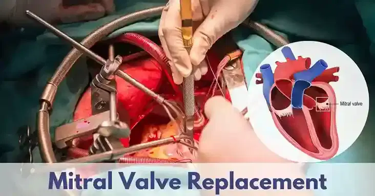
mitral valve surgery
5K+ International Patients Treated
40+ Source Countries Served
500+ Accredited Partner Hospitals
98% Patient Satisfaction
80% Average Savings vs USA
10K+ Doctors
NABH, JCI Accredited Hospitals
Free Treatment Plan
Free Consultation with Doctor
5+ Destinations Covered
About mitral valve surgery
Sources and Guidelines Referenced
This clinical guide is grounded in international clinical guidelines and seminal clinical studies, including: AHA/ACC Guideline for the Management of Patients With Valvular Heart Disease (Otto et al., 2020); ESC/EACTS Guidelines for the Management of Valvular Heart Disease (Vahanian et al., 2021); Society of Thoracic Surgeons (STS) Adult Cardiac Surgery Database Reports (2022); Heart Valve Society Clinical Practice Consensus Documents (2023); and landmark trials including the EVEREST II Trial (Feldman et al., 2011) and the COAPT Trial (Stone et al., 2018).
Mitral Valve Surgery: A Comprehensive Patient Guide
1. Definition and Medical Identity
Mitral valve surgery is a specialized open or minimally invasive cardiac operation designed to repair or replace a damaged mitral valve. The primary clinical objective is restoring forward blood flow, reducing left atrial pressure, and preserving left ventricular muscle function. It is categorized under adult cardiovascular surgery and structural heart interventions.
The mitral valve—named for its structural resemblance to a bishop's mitre—is one of four cardiac valves controlling blood circulation. Located on the left side of the heart, it acts as a precise one-way valve between the left atrium (upper collecting chamber) and the left ventricle (main pumping chamber). When the left ventricle contracts during systole (pumping phase), the mitral valve leaflets close tightly to prevent oxygen-rich blood from leaking backward into the lungs. When the valve becomes damaged by structural deterioration, infection, or disease, surgical intervention becomes necessary to restore normal heart function and protect the cardiovascular system.
2. The Underlying Condition or Need
Mitral valve surgery addresses structural breakdown of the valve leaflets, supporting cords, or tissue ring, which causes dangerous hemodynamic instability. Without intervention, untreated mitral valve disease forces the heart to overwork, ultimately causing progressive heart failure, irreversible ventricular enlargement, permanent atrial fibrillation, and severe pulmonary lung hypertension.
Pathology of the mitral valve typically presents in two distinct clinical forms, or a combination of both:
- Mitral regurgitation (MR): Incomplete closure of the valve leaflets allows blood to leak backward into the left atrium during ventricular contraction. Primary (degenerative) MR results from intrinsic physical damage to the valve leaflets or chordae tendineae (fibrous attachment cords). Secondary (functional) MR occurs when the left ventricle expands due to coronary artery disease or heart muscle weakness, stretching the valve ring (annulus) outward and pulling the leaflets apart.
- Mitral stenosis (MS): Structural narrowing and stiffening of the valve orifice restrict forward blood flow from the left atrium into the left ventricle during diastole (filling phase). This narrowing is most commonly caused by rheumatic heart disease, an autoimmune inflammatory reaction following untreated streptococcal throat infection, or heavy age-related calcification.
Patients typically experience progressive shortness of breath during physical activity (exertional dyspnea), fatigue, lower limb swelling (edema), persistent cough, and rapid or irregular heartbeats (palpitations). According to the AHA/ACC 2020 Guidelines, chronic pressure and volume overload lead to structural heart damage long before severe symptoms appear, making timely surgical evaluation essential.
3. How the Treatment Works — Mechanism
Mitral valve surgery restores dynamic blood flow by either reconstructive repair of the patient's native valve tissues or complete replacement using an artificial valve prosthesis. The underlying scientific principle relies on establishing fluid mechanical efficiency, eliminating backward leakage, and returning intra-cardiac blood pressures to normal physiological ranges.
During surgical repair, the operator mechanically corrects specific anatomical defects of the valve complex:
- Resection and reconstruction: Excess or floppy leaflet tissue causing valve collapse (prolapse) is precisely trimmed and reattached using ultra-fine sutures.
- Artificial chordae placement: Stretched or ruptured native cords are replaced with high-durability synthetic polytetrafluoroethylene (PTFE) sutures anchored to the internal papillary muscles.
- Annuloplasty ring insertion: A semi-rigid or flexible prosthetic ring is sewn onto the perimeter of the dilated valve base (annulus). This restores normal ring shape, brings the leaflets into close contact, and reinforces the repair long-term.
When native repair is not technically feasible due to extensive calcium accumulation or widespread structural destruction, complete replacement is performed. The surgeon removes the damaged leaflets while preserving subvalvular cords whenever possible to maintain left ventricular shape and pumping efficiency. A mechanical or biological valve prosthesis is then securely stitched directly into the valve orifice, re-establishing unidirectional fluid flow.
4. Types and Variations
Mitral valve surgery encompasses distinct operational strategies categorized by surgical approach (access route) and valve intervention technique (repair versus replacement). Clinicians determine the specific surgical strategy based on valve morphology, etiology, patient age, bleeding risk, and comorbidities as recommended by ESC/EACTS 2021 Guidelines.
The major surgical approaches and intervention types include:
| Surgical Method | Invasiveness | Key Technical Features | Primary Clinical Suitability |
|---|---|---|---|
| Open Sternotomy Repair | High (Full central chest incision) | Direct high-field visualization; full cardiac bypass exposure; gold-standard durability. | Complex multi-segment valve repair; concomitant aortic valve or coronary bypass surgery. |
| Minimally Invasive Thoracotomy (MIMVS) | Moderate (3-5 cm right chest incision) | Endoscopic camera visualization; peripheral femoral vessel bypass cannulation; less soft tissue trauma. | Isolated mitral valve repair or replacement; high priority on faster physical recovery. |
| Robotic-Assisted Valve Surgery | Low-Moderate (Small port incisions) | 3D high-definition magnification; wristed robotic instruments; superior precision in tight spaces. | Isolated degenerative mitral valve prolapse requiring precise complex leaflet repair. |
| Mechanical Valve Replacement | High (Sternotomy or Mini-thoracotomy) | Carbon bi-leaflet prosthetic replacement; lifetime structural durability; mandatory lifelong blood thinners. | Patients aged under 65 years without contraindications to long-term anticoagulation. |
| Bioprosthetic (Tissue) Replacement | High (Sternotomy or Mini-thoracotomy) | Bovine pericardial or porcine tissue valve; limited lifespan (12-15 years); no long-term blood thinners. | Patients aged over 65-70 years or those with high bleeding risks on anticoagulants. |
5. Who the Treatment Is For — Indications
Mitral valve surgery is indicated for individuals with severe structural mitral regurgitation or stenosis confirmed through comprehensive diagnostic imaging. Surgical intervention is recommended when valve malfunction threatens systemic cardiovascular stability or causes progressive heart muscle exhaustion, adhering to AHA/ACC and ESC clinical criteria.
Key clinical indications include:
- Symptomatic severe primary mitral regurgitation: Patients exhibiting exertion-induced shortness of breath, fatigue, or reduced exercise tolerance with severe primary MR (Class I recommendation).
- Asymptomatic severe primary mitral regurgitation with left ventricular dysfunction: Patients without symptoms whose left ventricular ejection fraction (LVEF) drops to 60% or lower, or whose left ventricular end-systolic diameter (LVESD) enlarges to 40 mm or greater (Class I recommendation).
- New-onset atrial fibrillation or pulmonary hypertension: Asymptomatic severe MR patients who develop secondary heart rhythm disturbances (atrial fibrillation) or elevated resting pulmonary artery systolic pressure greater than 50 mmHg (Class IIa recommendation).
- Severe symptomatic mitral stenosis: Patients with a mitral valve area (MVA) of 1.5 cm² or less experiencing functional limitations, particularly when non-surgical balloon valvuloplasty is anatomical contraindicated due to heavy calcification or left atrial blood clot.
- Infective endocarditis: Severe acute valve dysfunction caused by bacterial infection resulting in heart failure, persistent infection despite antibiotics, or large high-risk mobile vegetation.
6. Who the Treatment Is NOT For — Contraindications
Mitral valve surgery has definitive contraindications when the systemic risks of open heart surgery and cardiopulmonary bypass outweigh prospective survival or functional benefits. Clinicians evaluate individual surgical risk profiles using standardized scoring models from the Society of Thoracic Surgeons (STS).
Contraindications are stratified as follows:
- Absolute Contraindications:
- Severe irreversible left ventricular systolic failure (e.g., LVEF under 15-20%) where surgical trauma cannot produce recovery, unless performed alongside heart transplantation or mechanical circulatory support device insertion.
- Incurable advanced non-cardiac terminal illness with life expectancy under 12 months.
- Severe irreversible non-cardiac organ system failure, including end-stage liver cirrhosis or profound non-treatable neurological impairment.
- Relative Contraindications & Protocol Modifications:
- Prohibitive surgical risk profile secondary to severe frailty, host chest conditions (radiation-induced chest wall damage, severe porcelain aorta), or severe chronic obstructive pulmonary disease (COPD). These patients are referred for transcatheter alternatives.
- Active uncontrolled systemic infection or severe active bleeding disorder requiring emergency medical stabilization prior to elective valve reconstruction.
7. Alternatives and Clinical Comparison
Surgical mitral valve repair remains the standard of care for durable structural valve repair, but alternative medical and transcatheter approaches exist. Treatment decisions depend on surgical risk scoring, anatomical feasibility, and underlying etiology (primary versus secondary MR), as emphasized by the landmark EVEREST II and COAPT clinical trials.
The comparative clinical landscape is detailed below:
| Treatment Modality | Mechanism of Action | Level of Invasiveness | Durability & Key Trade-offs |
|---|---|---|---|
| Surgical Valve Repair | Direct anatomical restoration with annuloplasty ring and leaflet reconstruction. | Invasive (Sternotomy or Mini-thoracotomy under full bypass) | Gold-standard durability (>90% free from reoperation at 10-15 years); lowest long-term mortality; requires major surgical recovery. |
| Transcatheter Edge-to-Edge Repair (TEER / MitraClip) | Percutaneous mechanical clipping of anterior and posterior leaflets together via femoral vein. | Minimally Invasive Catheterization (No bypass, groin access) | Lower immediate procedural risk; rapidly shorter recovery; higher rate of residual regurgitation over long-term follow-up compared to surgery. |
| Optimal Medical Therapy (OMT) | Pharmaceutical unloading (ACE inhibitors, diuretics, ARNI, beta-blockers). | Non-invasive (Oral medications) | Manages secondary heart failure symptoms; fails to correct primary mechanical structural defect; disease continues to progress naturally. |
| Percutaneous Balloon Valvuloplasty | Transcatheter balloon inflation across stenotic valve to fracture fused commissures. | Minimally Invasive Catheterization | Effective non-surgical treatment exclusively for pliable, non-calcified rheumatic mitral stenosis; re-stenosis can occur over 5-10 years. |
8. Pre-Treatment Phase
The pre-treatment phase optimizes patient physiology, establishes precise anatomical mapping, and minimizes perioperative risk prior to entering the operating room. Comprehensive multidisciplinary planning involving heart team cardiologists, cardiac surgeons, and cardiac anesthesiologists ensures optimal treatment selection.
Key preoperative steps comprise:
- Diagnostic Workup and Anatomical Mapping: Patients undergo a high-resolution transesophageal echocardiogram (TEE) to generate three-dimensional images of leaflet mobility, tear locations, and annular dimensions. Coronary angiography (cardiac catheterization) is performed in patients over age 40 or those with cardiac risk factors to identify coexisting coronary artery blockages requiring simultaneous bypass grafting. A computed tomography (CT) chest scan evaluates aortic calcification and vascular access for minimally invasive planning.
- Systemic and Organ Optimization: Laboratory profiling includes complete blood counts, renal function panels, liver enzymes, coagulation studies, and blood typing. Pulmonary function testing (spirometry) assesses baseline lung capacity. Dental consultation and clearance are mandatory; any hidden tooth infections must be treated to prevent bacteria from migrating to the heart valve during surgery.
- Medication Management: Antiplatelet agents such as clopidogrel are typically discontinued 5 to 7 days before surgery to reduce bleeding risks, while essential heart medications like beta-blockers are maintained under specialist direction. Anticoagulant therapy (warfarin or direct oral anticoagulants) is held according to strict protocol timelines.
- Informed Consent and Preparation: Patients receive comprehensive education regarding surgical approach options, repair versus replacement likelihoods, biological versus mechanical valve trade-offs, anticipated ICU stays, and post-operative lifestyle adjustments.
9. The Procedure — Step-by-Step Clinical Detail
Mitral valve surgery is a highly coordinated procedure performed under general endotracheal anesthesia in a dedicated cardiac operating room. The operation requires specialized surgical support, advanced intraoperative TEE monitoring, and precise mechanical execution over three to five hours.
The intraoperative sequence progresses through distinct clinical steps:
Step 1: Anesthesia and Monitoring Setup
General endotracheal anesthesia is administered. Continuous invasive hemodynamic monitoring is established using an arterial line and a central venous catheter. A transesophageal echocardiography probe is positioned in the esophagus to capture real-time baseline heart images before incision.
Step 2: Surgical Access and Exposure
Surgical access is established either through a central median sternotomy (dividing the breastbone) or a right mini-thoracotomy (a 4-5 cm incision between the ribs on the right side of the chest). In robotic-assisted approaches, tiny keyhole camera and instrument ports are positioned across the right intercostal spaces.
Step 3: Cardiopulmonary Bypass Cannulation
The patient is connected to the cardiopulmonary bypass (heart-lung) machine. Cannulas are placed directly into the ascending aorta (or femoral artery) and the superior and inferior vena cavae (or femoral vein). Heparin blood thinner is administered to prevent clotting within the oxygenator circuit. Once full bypass support is established, the machine takes over oxygenating and circulating blood for the body.
Step 4: Cardiac Arrest and Protection
A cross-clamp is applied across the ascending aorta to isolate the heart. Cold, nutrient-dense cardioplegic solution is infused into the coronary arteries, causing the heart muscle to immediately relax and stop beating. This provides a still, dry, and protected surgical field.
Step 5: Valve Reconstructive Intervention
The left atrium is opened to reveal the mitral valve apparatus. The surgeon inspects the leaflets, chordae, and annulus under high magnification:
- If performing repair: Diseased tissue sections are resected, ruptured chordae are replaced with synthetic PTFE sutures, and a tailored annuloplasty ring is stitched into the valve perimeter to secure the correction.
- If performing replacement: Irreparably damaged leaflets are excised, preserving subvalvular tethering cords where possible. A mechanical or biological prosthetic valve sized precisely to the patient's heart is sutured securely along the fibrous ring.
Step 6: De-Airing, Re-Warming, and Weaning
The left atrial incision is closed. Air is meticulously evacuated from the heart chambers under continuous TEE guidance. The aortic cross-clamp is removed, allowing warm, oxygenated blood to flow back into the coronary arteries. The heart naturally resumes its rhythm or is electrically restored with a temporary pacemaker pulse. The patient is slowly weaned off the heart-lung machine as normal native circulation resumes.
Step 7: Hemostasis and Closure
Heparin is reversed using protamine sulfate to restore normal blood clotting. Temporary pacing wires and chest drainage tubes are placed around the heart to drain fluid. Sternal wires (for sternotomy) or rib-approximating sutures (for thoracotomy) are placed, followed by multi-layer closure of surgical incisions.
10. Immediate Post-Procedure Period
The immediate post-operative phase focuses on critical care stabilization, ventilator weaning, and hemodynamic monitoring. Patients are transferred directly from the operating suite to the Intensive Care Unit (ICU) under continuous mechanical support.
Primary milestones during the first 24 to 48 hours include:
- ICU Monitoring and Extubation: The patient remains sedated on a mechanical ventilator for the first 4 to 12 hours. Sedation is gradually reduced as body temperature normalizes and bleeding remains minimal. Once awake and breathing adequately, the breathing tube is removed. Hemodynamic parameters, heart rhythm, and chest tube drain outputs are continuously recorded.
- Pain Control Strategy: Pain management utilizes intravenous multi-modal regimens, combining non-opioid analgesics, patient-controlled analgesia (PCA) pumps, and local intercostal nerve blocks to facilitate deep breathing and coughing, preventing lung collapse (atelectasis).
- Early In-Bed and Bedside Mobilization: Within 18 to 24 hours post-procedure, physical therapy initiates basic seated positioning and standing bedside exercises. Incentive spirometry exercises encourage full lung expansion.
- Transfer to Step-Down Unit: Once chest drains are removed (typically post-operative day 1 or 2) and heart rhythm and blood pressure remain stable without continuous IV drip medications, the patient transitions from the ICU to a specialized step-down cardiac telemetry ward.
11. Recovery — Short and Long Term
Recovery following mitral valve surgery is a structured process spanning weeks to months. The total recovery duration varies based on the chosen surgical access route (sternotomy versus minimally invasive), overall health, and individual functional reserve.
The progressive recovery timeline moves through these phases:
- Weeks 1–2 (Hospital Discharge to Home): Hospital discharge typically occurs on post-operative days 5 to 7. Patients can walk short distances, manage personal hygiene, and navigate stairs slowly. Sternal precautions must be strictly observed for median sternotomy patients: avoid lifting, pushing, or pulling objects heavier than 5 to 10 pounds (2.3–4.5 kg), and refrain from driving due to airbag impact risks and delayed reaction times.
- Weeks 3–6 (Early Healing Phase): Surgical incisions heal fully. Energy levels gradually increase. Light activity and structured home walking programs expand daily. Driving is typically cleared around week 6 following clinical assessment and sternal stability confirmation. Patients with mechanical valves undergo blood testing to stabilize their oral anticoagulant dosing (INR target usually 2.5–3.5).
- Weeks 7–12 (Outpatient Cardiac Rehabilitation): Formal enrollment in an outpatient cardiac rehabilitation program provides supervised exercise training, nutritional education, and cardiovascular risk reduction. Most patients return to desk jobs or light work duties between weeks 6 and 8. Full return to heavy manual labor or intense athletic training occurs at 12 weeks upon physician clearance.
- Long-Term Surveillance: Follow-up echocardiograms are routinely performed at 1 to 3 months, 12 months, and annually thereafter to assess valve function, ventricular remodeling, and structural ring or prosthetic stability.
12. Risks, Side Effects, and Complications
While modern mitral valve surgery is highly safe when performed by experienced surgical teams, cardiac surgery carries inherent risks. Complication rates depend on age, pre-existing organ function, emergency status, and underlying cardiac pathology, as documented by the STS Adult Cardiac Surgery Database.
The comprehensive risk severity matrix is outlined below:
| Frequency Category | Clinical Complication / Side Effect | Incidence Rate | Management & Clinical Impact |
|---|---|---|---|
| Common / Mild | Postoperative Atrial Fibrillation (POAF) | 20% – 35% | Temporary irregular rhythm; managed with antiarrhythmic drugs and short-term anticoagulation; usually resolves within weeks. |
| Common / Mild | Incision site discomfort & mild localized edema | 15% – 25% | Managed with oral analgesics; self-limiting over 4-6 weeks as soft tissues heal. |
| Uncommon / Moderate | Postoperative Bleeding requiring reoperation | 2% – 5% | Requires surgical re-exploration to control bleeding vessels or clear pericardial blood clots. |
| Uncommon / Moderate | Acute Kidney Injury (AKI) | 2% – 5% | Transient renal impairment due to bypass circulation; managed with fluid optimization; temporary dialysis required in <1% of cases. |
| Rare / Serious | Ischemic Stroke or TIA | 1% – 2.5% | Caused by micro-thrombi or air emboli; minimized through thorough de-airing protocols and intraoperative TEE. |
| Rare / Serious | Complete Heart Block requiring permanent pacemaker | 2% – 4% | Disruption of electrical conduction near the mitral annulus; treated with permanent pacemaker implantation. |
| Rare / Serious | Deep Sternal Wound Infection / Mediastinitis | 1% | Severe tissue infection; requires long-term intravenous antibiotics and surgical wound debridement. |
| Rare / Serious | Operative Mortality (Elective cases) | 1% – 2% (Repair) 3% – 5% (Replacement) |
Risk stratified via STS score; heavily influenced by advanced patient age, emergent presentation, and organ failure. |
Patients must seek immediate emergency medical evaluation if they experience red-flag warning signs post-discharge: sudden severe shortness of breath, fever over 100.4°F (38°C), rapid or fluttering heartbeat, weight gain greater than 3 pounds in 24 hours, sudden weakness or speech changes, or pus-like drainage from surgical incisions.
13. Lifestyle and Behavioural Considerations
Long-term lifestyle modifications protect valve integrity, support heart muscle recovery, and lower future cardiovascular risk. Recommendations are tailored based on whether the patient underwent native repair or received a mechanical or biological prosthesis.
Key behavioral considerations include:
- Anticoagulation Compliance: Patients with mechanical prosthetic valves require lifelong oral anticoagulation (vitamin K antagonists such as warfarin) to prevent valve thrombosis. Regular blood monitoring of the International Normalized Ratio (INR) is required to keep levels within the therapeutic window (typically 2.5 to 3.5). Patients taking warfarin must maintain consistent dietary vitamin K intake (leafy greens). Patients with biological valves or successful repairs typically require antiplatelet therapy (aspirin) for 3 to 6 months unless concurrent atrial fibrillation dictates longer anticoagulation.
- Infective Endocarditis Prophylaxis: According to AHA guidelines, patients with artificial heart valves or those who have had valve repairs involving prosthetic rings or artificial cords must take prophylactic antibiotics prior to specific dental procedures to prevent bacterial bloodstream infection from colonizing the valve.
- Diet and Fluid Management: A heart-healthy, low-sodium diet (under 2,000 mg of sodium daily) prevents fluid retention and eases workloads on the recovering left ventricle. Fluid restrictions may be temporarily imposed during early recovery if heart failure symptoms persist.
- Physical Activity Progression: Continuous cardiovascular exercise, such as brisk walking, swimming, or cycling, is strongly recommended after full incision healing. Contact sports and heavy weightlifting must be discussed with a cardiologist, particularly for patients on lifelong blood thinners.
14. How Outcomes Are Measured
Surgical success is measured through physiological improvement, structural valve durability, symptom relief, and long-term survival metrics. Standardized reporting guidelines from the Society of Thoracic Surgeons and American College of Cardiology dictate post-operative assessment standards.
Primary outcome indicators include:
- Echocardiographic Endpoints: Immediate post-repair TEE in the operating room verifies valve competence. Successful repair is defined as residual regurgitation that is trace or mild (Grade 1 or less) without mean transvalvular gradient elevation (kept under 5 mmHg). Serial echocardiograms track left ventricular ejection fraction recovery and reverse chamber remodeling (reduction in enlarged atrial and ventricular volumes).
- Functional Class Improvement: Symptom relief is graded using the New York Heart Association (NYHA) Functional Classification. Over 90% of patients undergoing successful mitral repair move from symptomatic NYHA Class III/IV to asymptomatic or mildly symptomatic NYHA Class I/II within 6 months post-surgery.
- Durability and Freedom from Reoperation: Mitral valve repair demonstrates exceptional clinical durability. Long-term studies show that freedom from reoperation after primary degenerative valve repair exceeds 90% at 10 years and 80–85% at 20 years (David et al., JTCVS). For bioprosthetic replacements, valve degeneration typically accelerates after 10–15 years, eventually requiring re-intervention or transcatheter valve-in-valve replacement.
15. Recent Advances and Current Standard of Care
Over the past decade, mitral valve surgery has transitioned significantly toward minimally invasive execution, advanced imaging integration, and joint structural heart team management. Current standard of care emphasizes valve preservation (repair over replacement) whenever anatomically feasible.
Key technological and surgical advances include:
- Robotic and Endoscopic Instrumentation: High-definition 3D endoscopic cameras and wristed robotic instruments allow surgeons to perform complex leaflet reconstructions through 3-4 cm mini-thoracotomy incisions. Clinical registries demonstrate that robotic systems provide equivalent repair durability to traditional open sternotomy while reducing blood transfusion rates, wound infections, and length of hospital stay.
- Expanded Transcatheter Options (TEER and TMVR): Transcatheter edge-to-edge repair (MitraClip) has matured as a frontline treatment for secondary functional regurgitation in heart failure patients based on the COAPT trial results, as well as for high-risk surgical patients with primary MR. Dedicated transcatheter mitral valve replacement (TMVR) devices are undergoing active clinical trial evaluation for anatomies unsuited for clipping.
- Advanced Intraoperative Echocardiography: Real-time 3D transesophageal echocardiography allows surgical teams to view the valve from an "anatomical surgical perspective" before incision. This enables precise preoperative simulation and rapid intraoperative assessment of repair results.
16. Common Myths and Misconceptions
Misunderstandings regarding mitral valve surgery often lead to delayed presentation, unnecessary anxiety, or inappropriate treatment choices. Misconceptions can be addressed using established clinical evidence.
Common myths include:
Myth: Mitral valve replacement is always better and more definitive than repairing the native valve.
Reality: Clinical guidelines strongly favor mitral valve repair over replacement whenever possible. Repair preserves the patient's native heart tissue, maintains left ventricular function, carries lower operational mortality, eliminates the need for lifelong anticoagulation (in sinus rhythm), and yields superior long-term survival rates (Otto et al., AHA/ACC 2020).
Myth: Heart surgery cannot be safely performed without fully cutting open the breastbone.
Reality: Modern minimally invasive mitral valve surgery accesses the heart through a small incision between the ribs (mini-thoracotomy) or using robotic port access, leaving the breastbone completely intact while offering identical long-term repair quality.
Myth: Patients can wait to undergo surgery until severe symptoms severely restrict daily life.
Reality: Waiting for severe symptoms can cause permanent, irreversible damage to the left ventricular muscle and irreversible pulmonary hypertension. Guidelines recommend intervention at the onset of measurable heart enlargement or early muscle weakening, even if the patient feels completely asymptomatic.
Myth: Mechanical artificial valves are always superior to biological tissue valves because they never wear out.
Reality: While mechanical valves offer lifetime structural durability, they require strict lifelong blood thinners (warfarin) and daily blood testing, carrying ongoing bleeding risks. Biological tissue valves do not require long-term blood thinners, making them preferable for many older patients despite structural wear over 12 to 15 years.
Myth: Physical activity and regular exercise must be permanently avoided after heart valve surgery.
Reality: Following initial surgical healing and enrollment in cardiac rehabilitation, regular physical exercise is encouraged and beneficial for cardiovascular endurance and long-term functional recovery.
Myth: Catheter-based valve clips (MitraClip) have entirely replaced the need for traditional surgery.
Reality: Transcatheter repairs are valuable alternatives for high-risk surgical patients or specific heart failure populations. However, for surgical candidates with primary degenerative valve disease, surgical repair remains the gold standard, offering higher repair completeness and superior long-term durability.
17. Frequently Asked Questions
What is the overall survival rate after mitral valve surgery?
Elective mitral valve repair in stable patients carries an immediate operational survival rate exceeding 98-99% in high-volume cardiac surgical centers. Long-term life expectancy following successful valve repair frequently matches that of the age-matched general population, particularly when surgery is performed before severe left ventricular muscle damage occurs.
How long does a mitral valve repair last compared to a replacement?
A surgical mitral valve repair is highly durable, with over 90% of patients requiring no secondary operations at 10 to 15 years post-surgery. Biological tissue replacement valves typically last 12 to 15 years before structural deterioration occurs, whereas mechanical prosthetic valves last a lifetime structurally but require strict lifelong anticoagulation management.
How do surgeons decide between repairing or replacing the valve?
Surgeons evaluate valve morphology using preoperative 3D echocardiography. Repair is almost always preferred for degenerative disease (prolapse or tear) due to superior long-term survival and function. Replacement is performed when the valve tissue is irreparably destroyed by heavy calcification, severe rheumatic scarring, or active bacterial endocarditis.
What is the main difference between mechanical and tissue valves?
Mechanical valves are manufactured from durable pyrolytic carbon and last indefinitely, but require lifelong blood thinners (warfarin) to prevent blood clots. Tissue (bioprosthetic) valves, made from bovine or porcine heart tissue, do not require permanent blood thinners but naturally degrade over 12 to 15 years, potentially requiring future re-replacement.
How long will I spend in the intensive care unit after surgery?
Most patients remain in the intensive care unit (ICU) for 24 to 48 hours following surgery. During this window, critical care teams manage breathing tube removal, monitor heart rhythms, control blood pressure, and track chest tube drainage before transferring the patient to a step-down cardiac ward.
When can I safely resume driving after surgery?
Patients undergoing standard median sternotomy must refrain from driving for approximately 6 weeks to allow complete bone healing of the breastbone and reduce risk of chest trauma from seatbelts or airbags. Patients undergoing minimally invasive thoracotomy procedures may be cleared to drive earlier, typically within 3 to 4 weeks, depending on pain control and mobility.
Why is dental clearance required before mitral valve surgery?
Dental infections or active gum disease release bacteria directly into the bloodstream during dental manipulation. If bacteria attach to a newly repaired valve ring or prosthetic valve, it can cause endocarditis, a severe heart infection. Completing necessary dental treatments prior to surgery eliminates this risk factor.
Will I need to take blood thinners forever after surgery?
Lifelong blood thinners (warfarin) are required only if you receive a mechanical valve or have persistent underlying atrial fibrillation. Patients who undergo successful native valve repair or receive a bioprosthetic tissue valve typically take blood thinners for only 3 to 6 months post-surgery, switching later to daily aspirin if sinus rhythm is maintained.
What is atrial fibrillation, and why is it common after surgery?
Atrial fibrillation is a temporary irregular heartbeat originating in the top heart chambers. It occurs in 20% to 35% of cardiac surgery patients during the first few days post-procedure due to surgical manipulation, localized inflammation, and fluid shifts. It is manageable with medications and usually resolves during recovery.
How long does full recovery take after returning home?
While hospital discharge occurs within 5 to 7 days, full functional recovery takes 6 to 8 weeks for minimally invasive surgery and 8 to 12 weeks for open sternotomy. Energy levels, physical strength, and respiratory capacity gradually improve over this period with participation in outpatient cardiac rehabilitation.
Can mitral valve repair be repeated if it fails years later?
If a repaired mitral valve deteriorates after many years, re-operation can be performed to either re-repair or replace the valve. In select patients with prior ring annuloplasty or biological valve failure, minimally invasive transcatheter valve-in-valve catheter procedures may be considered as lower-risk alternatives to re-do surgery.
What lifting restrictions apply after cardiac surgery?
Patients who undergo median sternotomy must observe sternal precautions, restricting lifting, pushing, or pulling objects heavier than 5 to 10 pounds (2.3 to 4.5 kg) for 6 to 8 weeks. This restriction prevents motion across the healing breastbone, ensuring proper bone fusion and preventing wound breakdown.
Booking With DIVINHEAL
Get a free consultation to understand your treatment options
Cost Calculator
I know my treatment - show me cost from 3 hospitals
Plan My Journey
Tell us your condition and budget - our AI matches the right destination, hospital and doctor and visa pathway
Recommended Article

Booking With DIVINHEAL
Get a free consultation to understand your treatment options
Cost Calculator
I know my treatment - show me cost from 3 hospitals
Plan My Journey
Tell us your condition and budget - our AI matches the right destination, hospital and doctor and visa pathway
Recommended Article
Our Speciality and Treatments
oculoplastic surgery
Macular Degeneration Treatment (AMD)
Pediatric Ophthalmology
Squint / Strabismus Surgery (Adult)
ICL / Phakic IOL Implantation
LASIK / SMILE / PRK (Refractive Surgery)
Keratoconus Treatment (CXL / Rings)
Corneal Transplant (Keratoplasty)
Pediatric Squint Surgery
Diabetic Retinopathy Laser / Anti-VEGF Injections
Vitrectomy
Retinal Detachment Repair
Pterygium Surgery
Glaucoma Surgery (Trabeculectomy / MIGS)
Cataract Surgery (Phaco with IOL)






Meet Our Medical Specialists
Hospitals
NABH & JCI Accredited Hospitals in India,Turkey, Thailand & UAE.

Hisar Intercontinental Hospital
Saray Mah. Siteyolu Cad. No:7, Umraniye, 34768, Istanbul, Turkey

Medical Park Group, Istanbul
Fahrettin Kerim Gokay Cad. Tıbbiye Cd., Kadikoy, Istanbul, Turkey

Emsey Hospital, Pendik, Istanbul
Çamlık, Selçuklu Cd. No:22, 34912 Pendik/İstanbul, Türkiye
LIV Hospital, Istanbul
How DivinHeal Helps
We simplify your medical journey by providing comprehensive support and access to world-class healthcare.
Expert Specialist Matching
Connecting you with the world's top-rated medical experts.
Accredited Hospital Network
Access to JCI & NABH certified healthcare facilities.
Complete Travel Coordination
Hassle-free visa, stay, and local transport assistance.
24/7 Personal Care
Dedicated patient advisors supporting you at every step.
Journey Guidance
Full guidance from start to end of the patient treatment journey.
Expert Specialist Matching
Connecting you with the world's top-rated medical experts.
Everything you
need to know today
Browse through these common inquiries to better understand our patient-focused medical platform.
Yes, we work with a variety of insurance providers. Contact our team to verify your coverage.
Yes, we provide secure online consultations with experienced specialists.
Our care coordinators help match you with the most suitable specialist.
Absolutely. Your medical information is protected according to healthcare privacy standards.
Look at six things: accreditation (JCI or NABH), specialty depth, doctor credentials and experience, procedure-specific success rates, international patient support, and technology. DivinHeal's AI-driven matching evaluates every hospital in our accredited partner network on these dimensions and shortlists the best-fit options for your condition, budget, and country.
JCI (Joint Commission International) is the US-based global gold standard for hospital quality, recognised worldwide. NABH is India's national accreditation — accredited by ISQua, the same body that accredits JCI. Both signal independently verified safety and quality. Most of India's leading hospitals hold both.
Yes. All three welcome international patients through structured medical visa programs. India is the most established, treating patients from Africa, the Middle East, and South Asia at 60–80% lower cost. Thailand leads in cosmetic and dental care. The UAE is emerging in oncology and reproductive medicine.
Most patients save 50–80% on treatment costs. Heart bypass costs US $7,000–9,000 in India compared to $70,000–150,000 in the US. IVF costs $3,000–4,500 compared to $12,000–20,000 in the UK. Even after flights, visa, and accommodation, total savings remain 60–70%.
DivinHeal manages your entire non-medical journey: visa invitation letters, medical visa guidance, doctor appointments, teleconsultations, airport pickup, hospital-vetted accommodation for you and your attendant, language interpreters, local transport, cuisine preferences, and post-treatment follow-up — one dedicated coordinator from first enquiry to final follow-up.
You need a valid passport (6+ months validity), a medical visa (M-Visa for India — DivinHeal provides the hospital invitation letter), return flight tickets, recent medical reports and a doctor's referral, current prescription list, and proof of financial means. Any accompanying attendant needs their own passport and MX-Visa.
Still have more questions?
Book a call with our friendly team to learn how DivineHeal simplifies your healthcare journey.