Centres Of Excellence
Our Centres of Excellence bring together multidisciplinary teams to deliver precise diagnosis, advanced treatments, and superior outcomes across a wide spectrum of medical specialties.

OVERVIEW
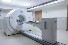
Radiology plays a pivotal role in healthcare, offering sophisticated tools for precise disease detection and treatment. From detailed anatomical imaging for early diagnosis and staging of cancers to delivering high-precision radiation therapy, the goal is to provide accurate insights for personalized patient care. DivinHeal connects patients with leading radiologists and radiation oncologists for advanced diagnostic and therapeutic procedures, ensuring comprehensive and integrated treatment plans.
PROCEDURE
Diagnostic Radiology procedures involve positioning the patient to capture images using X-rays, magnetic fields, or sound waves. Contrast agents may be administered orally or intravenously to enhance image clarity for specific organs. For Radiation Therapy, treatment planning involves detailed imaging (CT, MRI, PET) to precisely map the tumor and surrounding healthy tissues. A team of radiation oncologists, dosimetrists, and radiation therapists then designs a customized treatment plan, determining the exact dose and beam angles. During therapy, patients lie still on a treatment table while a linear accelerator delivers highly focused radiation beams to the target area, often over multiple daily sessions for several weeks. Modern techniques like IMRT and SBRT allow for highly conformal dose delivery, maximizing tumor kill while minimizing side effects.
BENEFITS
Radiology offers numerous benefits, from early and accurate disease diagnosis to highly targeted cancer treatment. Diagnostic imaging allows for non-invasive assessment of internal organs, reducing the need for exploratory surgeries. Radiation therapy provides a highly effective, often curative, treatment option for many cancers, preserving organ function where possible. Benefits include precise diagnosis, tailored treatment plans, reduced invasiveness, improved survival rates in cancer, and enhanced quality of life through effective disease management. DivinHeal focuses on connecting you with facilities offering these advanced benefits with a focus on patient safety and comfort.
RECOVERY
Recovery from Radiology procedures varies significantly based on the type of intervention. For diagnostic imaging (like X-rays, CT, MRI), recovery is typically immediate, allowing patients to resume normal activities almost immediately. For radiation therapy, recovery involves managing potential side effects, which can include fatigue, skin irritation, or localized discomfort, depending on the treated area. Most side effects are temporary and subside weeks or months after treatment completion. DivinHeal offers guidance on managing side effects, including dietary advice, skin care tips, and access to supportive therapies to ensure a smooth recovery and better quality of life post-treatment. Long-term wellness includes follow-up imaging and consultations.
WHAT WE TREAT
Radiology is instrumental in diagnosing and treating a wide array of conditions, including various forms of cancer (e.g., breast, lung, prostate, head and neck, gastrointestinal), benign tumors, cardiovascular diseases, neurological disorders, musculoskeletal injuries, and infectious diseases. Within the scope of cancer care, radiation therapy is effectively used to eradicate cancer cells, shrink tumors, or alleviate symptoms, either as a standalone treatment or in conjunction with surgery and chemotherapy.
PREPARATION
Preparation for Radiology procedures depends on the specific type. For most diagnostic scans, patients may be asked to fast for a few hours, remove metal objects, or change into a gown. If a contrast agent is used, a medical history regarding allergies or kidney function will be taken. For Radiation Therapy, preparation involves an initial consultation, detailed imaging for planning (simulation), and potentially a small tattoo or markers placed on the skin to ensure accurate daily positioning. Patients receive detailed instructions on diet, medications, and general well-being to optimize treatment outcomes and manage potential side effects.
RISKS
While largely safe, Radiology procedures carry some risks. Diagnostic imaging involves minimal radiation exposure, which is carefully managed to be 'as low as reasonably achievable' (ALARA principle). Contrast agents can cause allergic reactions in rare cases. For Radiation Therapy, risks include short-term side effects like fatigue, skin changes (redness, dryness), hair loss in the treated area, and localized discomfort or swelling. Long-term risks, though less common with modern precision techniques, can include tissue fibrosis, secondary cancers (very rare), or organ dysfunction, depending on the treated area and dosage. DivinHeal connects patients with experts who meticulously balance treatment efficacy with risk mitigation.
JOURNEY
Your Radiology treatment journey with DivinHeal begins with a thorough diagnostic phase, utilizing advanced imaging to accurately identify and stage your condition. This is followed by a multidisciplinary team consultation, where expert radiologists, oncologists, and other specialists collaborate to craft a personalized treatment plan, particularly for radiation therapy. Throughout your treatment, DivinHeal provides end-to-end coordination, including travel, accommodation, and continuous support, guiding you from initial consultation through the treatment sessions, recovery, and post-treatment follow-up, ensuring a seamless and reassuring experience.
OUTCOMES
The expected outcome of Radiology is a precise and timely diagnosis, providing clarity on your condition. This accurate information empowers you and your doctors to formulate the most effective treatment strategy, leading to improved health and well-being.
Related Links
Doctors For Treatment in Similar Locations
Best Hospital Near by for treatment
Related Treatments

Doppler Ultrasound
Treatment Price
$0.00 USDNo overview available
Treatment Overview
No overview available
Procedures
No procedure details available
Benefits
No benefits information available
Recovery Information
No recovery information available
CT Angiography
Treatment Price
$0.00 USDNo overview available
Treatment Overview
No overview available
Procedures
No procedure details available
Benefits
No benefits information available
Recovery Information
No recovery information available
Image-Guided Biopsy
Treatment Price
$0.00 USDNo overview available
Treatment Overview
No overview available
Procedures
No procedure details available
Benefits
No benefits information available
Recovery Information
No recovery information available
Vascular Embolisation
Treatment Price
$0.00 USDNo overview available
Treatment Overview
No overview available
Procedures
No procedure details available
Benefits
No benefits information available
Recovery Information
No recovery information available
PET-CT Scan
Treatment Price
$300.00 USDThe PET-CT Scan treatment leverages state-of-the-art nuclear medicine and radiography to visualize metabolic changes at the cellular level, often before structural changes are visible on traditional CT or MRI. It's a non-invasive procedure involving the injection of a small amount of a radioactive tracer (commonly FDG) to highlight areas of abnormal metabolic activity. This diagnostic insight is invaluable for cancer diagnosis, staging, recurrence detection, evaluating treatment response, and assessing various neurological and cardiac conditions.
Treatment Overview
The PET-CT Scan treatment leverages state-of-the-art nuclear medicine and radiography to visualize metabolic changes at the cellular level, often before structural changes are visible on traditional CT or MRI. It's a non-invasive procedure involving the injection of a small amount of a radioactive tracer (commonly FDG) to highlight areas of abnormal metabolic activity. This diagnostic insight is invaluable for cancer diagnosis, staging, recurrence detection, evaluating treatment response, and assessing various neurological and cardiac conditions.
Procedures
The PET-CT Scan procedure involves several steps. First, you will be asked to fast for a few hours prior to the scan. Upon arrival, a small amount of a radioactive tracer (most commonly FDG) is injected intravenously. You will then rest quietly for 45-90 minutes to allow the tracer to distribute throughout your body and accumulate in metabolically active cells. Following this uptake period, you will lie comfortably on a scanning bed that moves through the PET-CT scanner. The machine will capture images for approximately 20-30 minutes. During the scan, it's important to remain still. Once the imaging is complete, you can typically leave and resume normal activities.
Benefits
The PET-CT Scan offers numerous benefits, providing critical information that can significantly impact patient outcomes:<ul><li><strong>Comprehensive Diagnosis:</strong> Combines functional and anatomical imaging for a complete picture.</li><li><strong>Early Disease Detection:</strong> Can identify diseases like cancer at very early stages.</li><li><strong>Accurate Staging:</strong> Crucial for determining the extent of cancer spread, guiding PET-CT Scan diagnosis and therapy options.</li><li><strong>Effective Treatment Planning:</strong> Helps oncologists tailor radiation therapy and surgical plans precisely.</li><li><strong>Monitoring Treatment Response:</strong> Assesses how well chemotherapy and radiation therapy are working, allowing for timely adjustments.</li><li><strong>Recurrence Detection:</strong> Highly effective in identifying cancer recurrence.</li><li><strong>Non-Invasive:</strong> A comfortable procedure with minimal patient discomfort.</li><li><strong>Reduced Need for Biopsy:</strong> Can sometimes help differentiate benign from malignant lesions, potentially reducing the need for invasive biopsies.</li></ul>
Recovery Information
Recovery from a PET-CT Scan is typically straightforward as it is a non-invasive diagnostic procedure.<ul><li><h3>Immediately Post-Scan</h3><p>You can usually resume your normal activities immediately after the scan. It is recommended to drink plenty of fluids to help flush the small amount of radioactive tracer from your system.</p></li><li><h3>Tracer Clearance</h3><p>The radioactive tracer has a very short half-life and will naturally decay and leave your body within a few hours. No special isolation is required, though your doctor might advise limiting close contact with pregnant women or young children for a short period (typically 6-12 hours) as a precaution.</p></li><li><h3>Results and Follow-up</h3><p>The images will be interpreted by a nuclear medicine physician or radiologist, and a report will be sent to your referring doctor. Your doctor will then discuss the findings with you and plan any subsequent PET-CT Scan diagnosis and therapy options or monitoring. DivinHeal ensures timely delivery of your reports and facilitates communication with your healthcare team.</p></li></ul>
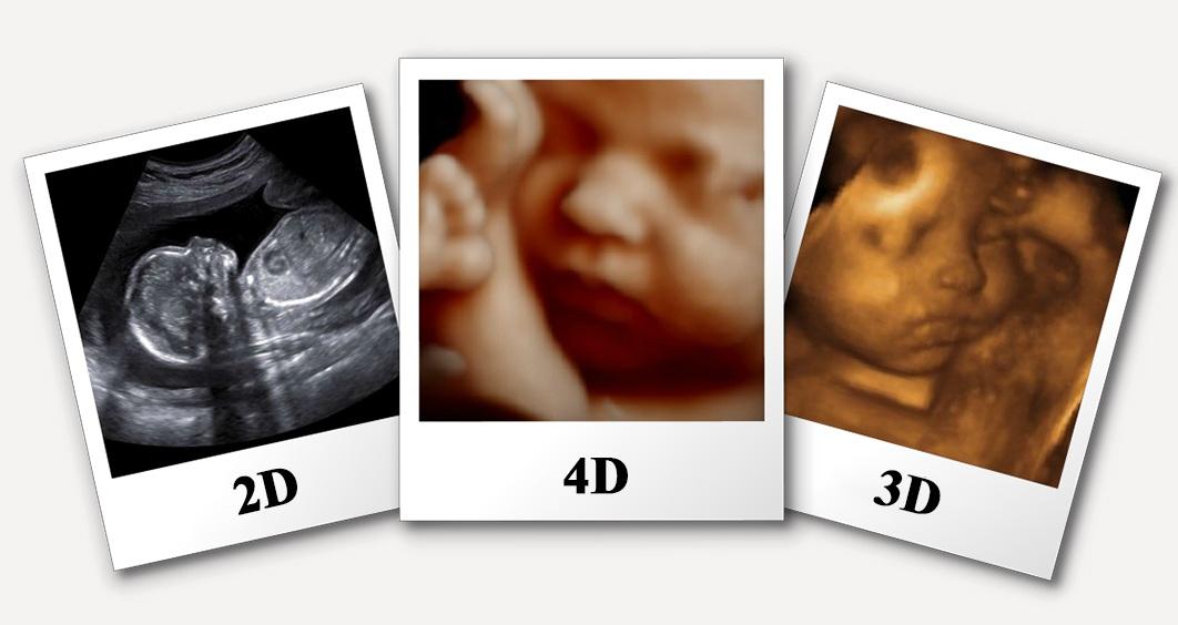
4D Ultrasound
Treatment Price
$80.00 USDThe primary goal of 4D Ultrasound is to provide highly detailed, dynamic images, offering greater diagnostic clarity and a unique interactive experience, particularly for expectant parents. This technology enhances the assessment of fetal development, maternal health, and various internal organ structures with superior spatial and temporal resolution.
Treatment Overview
The primary goal of 4D Ultrasound is to provide highly detailed, dynamic images, offering greater diagnostic clarity and a unique interactive experience, particularly for expectant parents. This technology enhances the assessment of fetal development, maternal health, and various internal organ structures with superior spatial and temporal resolution.
Procedures
The 4D Ultrasound procedure is a comfortable, non-invasive process. The patient will be asked to lie comfortably on an examination table, typically on their back. A clear, water-based, hypoallergenic gel is applied to the skin over the area of the body to be scanned (e.g., the abdomen for prenatal scans, the pelvic region for gynecological examinations, or specific organ areas). The sonographer, a highly trained technologist, then gently moves a handheld device called a transducer over the gelled skin. This transducer emits high-frequency sound waves that travel into the body and bounce off internal structures, creating echoes. These echoes are captured by the transducer and instantaneously transmitted to a sophisticated computer system, which processes them into live, moving three-dimensional images displayed on a monitor. The sonographer will carefully capture various views and take necessary measurements. Patients may often be able to observe the real-time images during the scan. The entire procedure is painless and generally lasts between 20 to 45 minutes, depending on the specific area being examined and the level of detailed visualization required.
Benefits
<ul><li><strong>Enhanced Diagnostic Accuracy:</strong> Provides dynamic, three-dimensional views, offering superior detail for detecting subtle anomalies or understanding complex anatomies that might be missed in 2D scans.</li><li><strong>Real-time Visualization:</strong> Allows for immediate observation of movements, blood flow, and organ function, which is crucial for dynamic assessments, particularly in fetal studies and cardiac evaluations.</li><li><strong>Non-invasive and Safe:</strong> Utilizes high-frequency sound waves rather than ionizing radiation, making it completely safe for all patient populations, including pregnant women and fetuses, with no known harmful side effects.</li><li><strong>Improved Patient Experience & Bonding:</strong> Offers a profound emotional experience for expectant parents, allowing them to see their baby's live movements and expressions, fostering an early connection.</li><li><strong>Better Pre-Procedural Planning:</strong> More detailed and dynamic pre-procedure imaging can aid specialists in planning subsequent interventions or treatments if required, leading to better outcomes.</li><li><strong>Cost-Effective Advanced Imaging:</strong> Especially in destinations facilitated by DivinHeal like India, 4D Ultrasound offers high-definition, state-of-the-art imaging at a fraction of the cost found in many Western countries, without compromising quality.</li></ul>
Recovery Information
<p>Recovery from a 4D Ultrasound is immediate as it is a completely non-invasive diagnostic procedure. There is no downtime, and patients can seamlessly resume all normal daily activities immediately after the scan. No special post-procedure care, restrictions, or medications are typically needed. Patients simply receive their diagnostic report and images, and then proceed to consult with their healthcare provider to discuss the findings and any subsequent steps. For expectant parents, the experience often leaves a lasting memory of seeing their baby in motion, with no physical recovery required.</p>
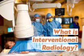
Interventional Radiology Procedures
Treatment Price
$2500.00 USDInterventional Radiology Procedures focus on precision and minimal invasiveness. Utilizing advanced imaging modalities such as X-ray, ultrasound, CT, and MRI, interventional radiologists guide small catheters, wires, or needles through tiny incisions to access and treat diseases directly at their source. The primary goals include reducing patient discomfort, minimizing recovery time, and achieving effective therapeutic results while preserving healthy tissue. DivinHeal connects you to world-class facilities offering these advanced procedures.
Treatment Overview
Interventional Radiology Procedures focus on precision and minimal invasiveness. Utilizing advanced imaging modalities such as X-ray, ultrasound, CT, and MRI, interventional radiologists guide small catheters, wires, or needles through tiny incisions to access and treat diseases directly at their source. The primary goals include reducing patient discomfort, minimizing recovery time, and achieving effective therapeutic results while preserving healthy tissue. DivinHeal connects you to world-class facilities offering these advanced procedures.
Procedures
Interventional Radiology Procedures typically begin with an initial consultation and thorough diagnostic imaging (CT, MRI, ultrasound). On the day of the procedure, you will be prepared by the medical team. Local anesthesia is usually administered at the skin entry point to minimize discomfort. Using real-time imaging guidance, the interventional radiologist makes a small incision, often just a few millimeters, and inserts a thin catheter or needle. This instrument is carefully guided to the target area within the body. Once in place, specialized tools are used to perform the intervention, such as delivering medication, blocking blood vessels (embolization), destroying tumors (ablation), opening narrowed vessels (angioplasty and stenting), or taking a biopsy. Upon completion, the instruments are removed, and the small incision is closed, usually with a small bandage. The entire process is meticulously monitored to ensure patient safety and procedural success.
Benefits
<h2>Benefits of Interventional Radiology Procedures</h2><ul><li><strong>Minimally Invasive:</strong> Performed through tiny incisions, leading to less pain and discomfort.</li><li><strong>Faster Recovery:</strong> Shorter hospital stays and quicker return to daily activities compared to traditional surgery.</li><li><strong>Reduced Risk:</strong> Lower risk of complications such as infection, bleeding, and scarring.</li><li><strong>Precision and Accuracy:</strong> Real-time imaging guidance ensures highly targeted treatment.</li><li><strong>Outpatient Potential:</strong> Many procedures can be performed on an outpatient basis.</li><li><strong>Preservation of Function:</strong> Often spares healthy tissue, minimizing impact on organ function.</li><li><strong>Effective Alternative:</strong> Provides viable treatment options for patients unsuitable for open surgery.</li><li><strong>Cost-Effective:</strong> Often more affordable than traditional surgical interventions, especially in destinations like India.</li></ul>
Recovery Information
<h2>Interventional Radiology Procedures Recovery Time and Tips</h2><p>Recovery from Interventional Radiology Procedures is generally much faster and less complicated than from open surgery. Most patients experience a rapid return to normal activities, though specific timelines vary based on the procedure's complexity and individual health factors.</p><h3>Typical Recovery Timeline</h3><ul><li><strong>Immediate Post-Procedure:</strong> Patients are usually monitored for a few hours in a recovery area. Some procedures may require an overnight stay (1-3 days).</li><li><strong>First Few Days:</strong> Mild discomfort, bruising, or swelling at the incision site is common and can be managed with over-the-counter pain relievers.</li><li><strong>First Week:</strong> Most patients can resume light activities. Strenuous exercise, heavy lifting, and prolonged standing may be restricted.</li><li><strong>2-4 Weeks:</strong> Full recovery and return to most normal activities, depending on the procedure. For more complex interventions like tumor ablations, full recovery might extend to several weeks.</li></ul><h3>Recovery Tips for Interventional Radiology Procedures</h3><ul><li><strong>Follow Post-Procedure Instructions:</strong> Adhere strictly to your interventional radiologist's specific advice regarding wound care, medication, and activity restrictions.</li><li><strong>Manage Pain:</strong> Use prescribed or recommended pain relief as needed.</li><li><strong>Stay Hydrated:</strong> Drink plenty of fluids to aid recovery and help flush contrast dye from your system (if used).</li><li><strong>Rest Adequately:</strong> Allow your body time to heal. Avoid overexertion.</li><li><strong>Monitor Incision Site:</strong> Watch for any signs of infection, such as increased redness, swelling, pus, or fever, and report them immediately to your care team.</li><li><strong>Light Activity:</strong> Gradually reintroduce physical activity as advised by your doctor. Walking is often encouraged soon after.</li><li><strong>Nutritious Diet:</strong> Eat a balanced diet to support healing.</li><li><strong>Emotional Support:</strong> DivinHeal provides access to counseling and support groups to help manage any emotional aspects of recovery.</li></ul>

3 Tesla MRI Scan
Treatment Price
$300.00 USDThe primary goal of a 3 Tesla MRI scan is to provide unparalleled diagnostic insights into complex medical conditions, particularly in neurology, orthopedics, cardiology, and oncology. By generating very strong magnetic fields and radio waves, it creates detailed cross-sectional images, enabling precise detection of abnormalities, tumors, injuries, and degenerative diseases. DivinHeal connects patients with leading diagnostic centers equipped with state-of-the-art 3T MRI technology for accurate and timely assessments.
Treatment Overview
The primary goal of a 3 Tesla MRI scan is to provide unparalleled diagnostic insights into complex medical conditions, particularly in neurology, orthopedics, cardiology, and oncology. By generating very strong magnetic fields and radio waves, it creates detailed cross-sectional images, enabling precise detection of abnormalities, tumors, injuries, and degenerative diseases. DivinHeal connects patients with leading diagnostic centers equipped with state-of-the-art 3T MRI technology for accurate and timely assessments.
Procedures
The 3 Tesla MRI scan involves several steps: 1. **Preparation**: Patients remove all metallic objects, jewelry, and often change into a gown. Inform the technician about any implants or claustrophobia. 2. **Positioning**: The patient lies on a comfortable, movable table that slides into the MRI machine's tunnel. The body part to be scanned is positioned correctly, sometimes with a specialized coil. 3. **Contrast Administration (if needed)**: An intravenous contrast agent may be injected, typically Gadolinium, to enhance visibility of certain tissues or pathologies. 4. **Scanning**: The machine generates a powerful magnetic field and emits radio waves. Patients hear loud knocking or thumping noises; earplugs or headphones are provided. It's crucial to remain perfectly still during this process to avoid blurring images. 5. **Duration**: The scan duration varies based on the body part and complexity, typically ranging from 30 minutes to an hour. 6. **Completion**: Once the scan is complete, the patient is removed from the machine and can usually leave immediately. 7. **Image Interpretation**: Radiologists meticulously analyze the images to provide a comprehensive diagnostic report to the referring physician.
Benefits
The benefits of undergoing a 3 Tesla MRI scan are significant, offering superior diagnostic capabilities:<ul><li>**Exceptional Image Clarity**: Produces incredibly detailed, high-resolution images, aiding in the detection of even minute abnormalities.</li><li>**Enhanced Diagnostic Accuracy**: The increased signal strength allows for more precise identification of lesions, tumors, inflammation, and structural changes.</li><li>**Faster Scan Times**: Often reduces the time patients need to spend inside the scanner, improving comfort.</li><li>**Better for Subtle Conditions**: Particularly effective for diagnosing complex neurological conditions, small joint injuries, and early-stage diseases.</li><li>**Non-Invasive and Radiation-Free**: Uses magnetic fields and radio waves instead of ionizing radiation, making it a safe option for repeated scans.</li><li>**Versatile Applications**: Useful across a broad spectrum of medical specialties, from brain and spine to abdomen and extremities.</li><li>**Improved Patient Experience**: While still requiring patients to remain still, faster scans can reduce anxiety for some.</li></ul>
Recovery Information
There is typically no recovery period required after a 3 Tesla MRI scan, as it is a non-invasive diagnostic procedure. Patients can usually resume their normal activities immediately following the scan. If a contrast agent was administered, minor side effects like a temporary metallic taste or mild nausea are rare but possible; staying hydrated helps. Our team ensures you receive clear instructions and your diagnostic reports are processed efficiently for prompt review by your consulting physician.
Interventional Radiology: UFE (Fibroids)
Treatment Price
$3000.00 USDUFE offers a non-surgical alternative to hysterectomy or myomectomy for women suffering from symptomatic uterine fibroids. The primary goal of UFE is to significantly reduce fibroid size and associated symptoms, improving quality of life while preserving the uterus. The procedure involves guiding a catheter through an artery to the uterus and injecting embolic agents to cut off blood flow to the fibroids.
Treatment Overview
UFE offers a non-surgical alternative to hysterectomy or myomectomy for women suffering from symptomatic uterine fibroids. The primary goal of UFE is to significantly reduce fibroid size and associated symptoms, improving quality of life while preserving the uterus. The procedure involves guiding a catheter through an artery to the uterus and injecting embolic agents to cut off blood flow to the fibroids.
Procedures
Uterine Fibroid Embolization (UFE) is performed by an interventional radiologist in an angiography suite. The patient receives local anesthesia and conscious sedation. A small incision (2-3 mm) is made, typically in the groin, to access the femoral artery. A thin catheter is then guided under X-ray (fluoroscopic) guidance through the arterial system to locate the uterine arteries supplying the fibroids. Once the catheter is in position, microscopic particles (embolic agents) are injected into these arteries. These particles block the blood flow to the fibroids, causing them to shrink. The procedure is usually completed within 1-2 hours, and the catheter is then removed.
Benefits
<h2>Benefits of Uterine Fibroid Embolization (UFE)</h2><ul><li><h3>Minimally Invasive Procedure</h3><p>UFE requires only a tiny incision, typically in the groin or wrist, significantly reducing pain, scarring, and risk of infection compared to open surgery.</p></li><li><h3>Uterus Preservation</h3><p>Unlike hysterectomy, UFE allows women to retain their uterus, a significant advantage for those who wish to avoid surgical removal of an organ or desire future pregnancy (though individual fertility outcomes vary).</p></li><li><h3>Faster Recovery Time</h3><p>Patients typically experience a quicker return to normal activities, with most resuming light activities within a few days to a week, compared to several weeks for surgical alternatives.</p></li><li><h3>Effective Symptom Relief</h3><p>UFE boasts a high success rate in alleviating heavy menstrual bleeding, pelvic pain, and pressure symptoms caused by fibroids, with significant improvement reported by a majority of patients.</p></li><li><h3>Global Accessibility & Affordability</h3><p>Through DivinHeal, access world-class UFE treatment in India at a fraction of the cost found in Western countries, ensuring quality care is accessible to all.</p></li></ul>
Recovery Information
<h2>Recovery and Life After UFE (Fibroids) Treatment</h2><h3>Immediate Post-Procedure Care</h3><p>After UFE, patients typically spend a few hours to an overnight stay in the hospital for observation and pain management. It's common to experience pelvic pain or cramping, known as 'Post-Embolization Syndrome,' which can be managed effectively with medication. Nausea, low-grade fever, and fatigue are also possible. DivinHeal ensures comprehensive pain management and monitoring during this phase.</p><h3>Short-Term Recovery (Days to Weeks)</h3><p>Most patients can return to light activities within 3-5 days and resume normal daily routines within 1-2 weeks. Strenuous exercise should be avoided for at least a week. Follow-up appointments are scheduled to monitor recovery and fibroid shrinkage. DivinHeal's care coordinators will assist with all follow-up arrangements, ensuring continuous support.</p><h3>Long-Term Outlook</h3><p>Fibroids continue to shrink over the next few months, with maximal shrinkage typically observed at 3-6 months. Symptoms like heavy bleeding and pain generally improve significantly within the first few menstrual cycles. Long-term follow-up with your gynaecologist and interventional radiologist is recommended, often including follow-up MRI scans to assess fibroid size and uterine health. DivinHeal emphasizes a holistic approach to recovery, including guidance on nutrition, physical activity, and emotional well-being to support a full and sustained return to health.</p>
Interventional Radiology: TACE / Y-90 (Liver Tumors)
Treatment Price
$5000.00 USDOverview of TACE and Y-90 Treatments for Liver Tumors
Targeted Liver Cancer Therapies
TACE and Y-90 (Radioembolization) are sophisticated, image-guided procedures used to treat various liver tumors, including hepatocellular carcinoma (HCC) and metastatic cancers. These therapies aim to deliver high doses of therapeutic agents directly to the tumor while minimizing systemic exposure, maximizing effectiveness, and preserving liver function.
How They Work
Both procedures involve inserting a catheter into the femoral artery and guiding it to the hepatic artery supplying the tumor. TACE delivers a mixture of chemotherapy drugs and embolic agents to block the tumor's blood supply. Y-90 employs microscopic radioactive spheres that lodge in the tumor vasculature, emitting localized radiation.
Treatment Overview
<h2>Overview of TACE and Y-90 Treatments for Liver Tumors</h2><ul><li><h3>Targeted Liver Cancer Therapies</h3><p>TACE and Y-90 (Radioembolization) are sophisticated, image-guided procedures used to treat various liver tumors, including hepatocellular carcinoma (HCC) and metastatic cancers. These therapies aim to deliver high doses of therapeutic agents directly to the tumor while minimizing systemic exposure, maximizing effectiveness, and preserving liver function.</p></li><li><h3>How They Work</h3><p>Both procedures involve inserting a catheter into the femoral artery and guiding it to the hepatic artery supplying the tumor. TACE delivers a mixture of chemotherapy drugs and embolic agents to block the tumor's blood supply. Y-90 employs microscopic radioactive spheres that lodge in the tumor vasculature, emitting localized radiation.</p></li></ul>
Procedures
Before the Procedure: You will undergo detailed imaging (CT, MRI) and blood tests to map the liver's blood supply and assess tumor characteristics. A pre-procedure angiography might be performed to plan the best approach. You will be asked to fast for several hours prior to the procedure. During TACE / Y-90: The procedure is typically performed in an angiography suite under conscious sedation. A small incision is made in the groin to access the femoral artery. A thin catheter is then guided through the arterial system, under real-time X-ray guidance, to the specific hepatic artery branches feeding the liver tumor(s). For TACE, a mixture of chemotherapy drugs and embolic agents is injected directly into these arteries. For Y-90, radioactive microspheres are injected. The entire process can take 1-3 hours. After the Procedure: The catheter is removed, and pressure is applied to the groin site to prevent bleeding. You will be monitored in a recovery area for several hours, then transferred to a hospital room for observation, typically for 1-2 days.
Benefits
<h2>Benefits of TACE and Y-90 for Liver Tumors</h2><ul><li><h3>Targeted Efficacy</h3><p>Both TACE and Y-90 deliver therapeutic agents directly to the tumor, minimizing systemic side effects common with traditional chemotherapy or external beam radiation. This precision maximizes tumor control.</p></li><li><h3>Minimally Invasive</h3><p>Performed through a small incision, these procedures avoid major surgery, resulting in less pain, shorter hospital stays, and quicker recovery times compared to open surgical options.</p></li><li><h3>Preservation of Healthy Liver Tissue</h3><p>By selectively targeting the tumor's blood supply or delivering localized radiation, TACE and Y-90 spare much of the healthy liver tissue, maintaining overall liver function, which is crucial for patients with compromised liver health.</p></li><li><h3>Improved Quality of Life & Survival</h3><p>For patients with unresectable liver tumors, these therapies can significantly slow tumor progression, reduce tumor size, alleviate symptoms, improve quality of life, and often extend survival, providing renewed hope.</p></li><li><h3>Suitable for Non-Surgical Candidates</h3><p>TACE and Y-90 are vital options for patients who are not candidates for surgery due to tumor location, size, number, or underlying health conditions.</p></li></ul>
Recovery Information
<h2>Recovery After TACE / Y-90 Treatment for Liver Tumors</h2><ul><li><h3>Immediate Post-Procedure</h3><p>Patients typically stay in the hospital for 1-2 days for observation. Common immediate side effects, often referred to as "post-embolization syndrome," can include mild fever, pain in the abdomen (managed with medication), and nausea/vomiting. These usually subside within a few days.</p></li><li><h3>At-Home Recovery</h3><p>Most patients can return to light activities within 1-2 weeks. It's crucial to follow your doctor's instructions regarding activity restrictions, diet, and medication. Fatigue is common and may persist for several weeks. Adequate rest and hydration are key to a smooth recovery.</p></li><li><h3>Long-Term Monitoring & Lifestyle</h3><p>Regular follow-up appointments, including imaging scans (CT, MRI), are essential to monitor the tumor's response to treatment and overall liver health. A healthy lifestyle, including a balanced diet and avoiding alcohol, supports liver function and overall well-being. DivinHeal offers resources for nutritional guidance and emotional support during your long-term recovery journey.</p></li></ul>
Digital Mammography
Treatment Price
$75.00 USDDigital Mammography aims to detect breast changes, such as calcifications or masses, which could indicate breast cancer. Using digital detectors instead of film, it allows for enhanced image manipulation, storage, and transfer, leading to more accurate diagnoses and often lower radiation doses compared to traditional mammography. It's a cornerstone of preventative health.
Treatment Overview
Digital Mammography aims to detect breast changes, such as calcifications or masses, which could indicate breast cancer. Using digital detectors instead of film, it allows for enhanced image manipulation, storage, and transfer, leading to more accurate diagnoses and often lower radiation doses compared to traditional mammography. It's a cornerstone of preventative health.
Procedures
A Digital Mammography procedure typically involves the following steps: 1. You will be asked to remove clothing and jewelry from the waist up and wear a gown. 2. A trained mammography technologist will position one of your breasts on a special platform. 3. Compression is applied to flatten the breast, which helps to spread the tissue, reduce radiation dose, and prevent motion blur. While this might cause brief discomfort, it's essential for clear images. 4. X-ray images are taken from different angles (usually two views per breast). 5. The digital images are immediately available for review on a computer monitor by the technologist and later by a radiologist. The entire process for both breasts usually takes about 15-30 minutes.
Benefits
<h2>Benefits of Digital Mammography</h2><ul><li><h3>Early Breast Cancer Detection</h3><p>The most significant benefit is its ability to detect breast cancer at its earliest, most treatable stages, often before any symptoms are present.</p></li><li><h3>Improved Accuracy</h3><p>Digital imaging offers superior contrast and detail, making it easier for radiologists to identify subtle changes, especially in women with dense breasts.</p></li><li><h3>Reduced Radiation Exposure</h3><p>Many digital systems use lower radiation doses compared to traditional film mammography.</p></li><li><h3>Faster & More Efficient</h3><p>Digital images are acquired and processed quickly, leading to shorter appointment times and immediate review by radiologists.</p></li><li><h3>Enhanced Diagnostic Capabilities</h3><p>Radiologists can manipulate images (zoom, adjust contrast) to get a clearer view of suspicious areas, reducing the need for repeat scans.</p></li><li><h3>Peace of Mind</h3><p>Regular screening provides peace of mind, knowing that you are actively monitoring your breast health and taking proactive steps for early intervention.</p></li></ul>
Recovery Information
<h2>Digital Mammography: Minimal Recovery, Maximum Peace of Mind</h2><p>Digital Mammography is a quick, outpatient diagnostic procedure with virtually no recovery time. You can resume your normal daily activities immediately after your appointment. Any mild discomfort from breast compression during the scan typically subsides within minutes. The main "recovery" aspect is often the wait for results and any subsequent follow-up actions if an abnormality is detected. DivinHeal ensures prompt result delivery and seamless coordination for any necessary next steps, providing emotional support and expert guidance throughout.</p>
DEXA Scan (Bone Density)
Treatment Price
$80.00 USDOverview of DEXA Scan (Bone Density)
The DEXA Scan is the gold standard for bone mineral density measurement, crucial for the early detection and management of conditions like osteoporosis and osteopenia. This quick, safe procedure helps physicians assess bone strength and fracture risk. DivinHeal facilitates access to state-of-the-art diagnostic centers globally, ensuring you receive accurate and timely results for your bone health journey. We focus on providing quality care and transparency for every DEXA Scan (Bone Density) diagnosis and therapy options.
Treatment Overview
<h2>Overview of DEXA Scan (Bone Density)</h2><p>The DEXA Scan is the gold standard for bone mineral density measurement, crucial for the early detection and management of conditions like osteoporosis and osteopenia. This quick, safe procedure helps physicians assess bone strength and fracture risk. DivinHeal facilitates access to state-of-the-art diagnostic centers globally, ensuring you receive accurate and timely results for your bone health journey. We focus on providing quality care and transparency for every DEXA Scan (Bone Density) diagnosis and therapy options.</p>
Procedures
The patient lies still on a padded table. A scanner arm moves slowly over the target areas, typically the lower spine and hips. Low-dose X-rays are emitted and absorbed by the bones and soft tissues. A detector measures the X-rays that pass through, and a computer uses this data to calculate bone mineral density. The process is quick, painless, and non-invasive, usually taking 10-20 minutes.
Benefits
<h2>Benefits of a DEXA Scan (Bone Density)</h2><ul><li><h3>Accurate Diagnosis</h3><p>The gold standard for precise measurement of bone mineral density, allowing for early detection of osteoporosis and osteopenia.</p></li><li><h3>Fracture Risk Assessment</h3><p>Helps predict future fracture risk, enabling proactive preventative measures.</p></li><li><h3>Non-Invasive and Painless</h3><p>A comfortable procedure requiring no injections or discomfort.</p></li><li><h3>Low Radiation Exposure</h3><p>Uses very low doses of radiation, making it a safe diagnostic option.</p></li><li><h3>Monitors Treatment Effectiveness</h3><p>Allows physicians to track changes in bone density over time and evaluate the success rate of interventions.</p></li><li><h3>Guides Personalized Treatment</h3><p>Provides crucial data for developing individualized bone density treatment plans and hormone therapy, if needed.</p></li></ul>
Recovery Information
<h2>Understanding Your DEXA Results and Next Steps for Bone Health</h2><p>A DEXA scan itself requires no recovery time, and you can resume normal activities immediately. However, the 'recovery' aspect shifts to understanding your results and implementing recommended strategies for bone health. Your DEXA report will include T-scores and Z-scores, which your physician will interpret to determine your bone density status.<ul><li><b>Normal Bone Density:</b> T-score of -1.0 or above.</li><li><b>Osteopenia (Low Bone Mass):</b> T-score between -1.0 and -2.5.</li><li><b>Osteoporosis:</b> T-score of -2.5 or below.</li></ul>Depending on your results, your doctor might recommend lifestyle modifications (diet, exercise), calcium and Vitamin D supplementation, or specific medications (e.g., bisphosphonates, hormone therapy for osteoporosis). DivinHeal assists in facilitating these follow-up consultations and provides resources for long-term bone health management, ensuring you get the most out of your DEXA Scan (Bone Density) diagnosis and therapy options for optimal well-being.</p>
CT Scan (Contrast / Non-contrast)
Treatment Price
$250.00 USDOverview of CT Scan (Contrast / Non-contrast)
The primary goal of a CT Scan is to provide highly detailed anatomical information that cannot be obtained from standard X-rays. This diagnostic precision is crucial for:
Accurate Diagnosis:
Identifying tumors, fractures, internal bleeding, and organ damage.Disease Staging:
Determining the extent of diseases like cancer, which is vital for treatment planning.Treatment Planning:
Guiding biopsies, surgeries, radiation therapy, and other interventional procedures.Monitoring Treatment Response:
Assessing the effectiveness of therapies over time.
DivinHeal ensures access to state-of-the-art CT technology and expert radiologists, guaranteeing precise diagnostics critical for effective medical management.
Treatment Overview
<h2>Overview of CT Scan (Contrast / Non-contrast)</h2><p>The primary goal of a CT Scan is to provide highly detailed anatomical information that cannot be obtained from standard X-rays. This diagnostic precision is crucial for:</p><ul> <li><h3>Accurate Diagnosis:</h3> Identifying tumors, fractures, internal bleeding, and organ damage.</li> <li><h3>Disease Staging:</h3> Determining the extent of diseases like cancer, which is vital for treatment planning.</li> <li><h3>Treatment Planning:</h3> Guiding biopsies, surgeries, radiation therapy, and other interventional procedures.</li> <li><h3>Monitoring Treatment Response:</h3> Assessing the effectiveness of therapies over time.</li></ul><p>DivinHeal ensures access to state-of-the-art CT technology and expert radiologists, guaranteeing precise diagnostics critical for effective medical management.</p>
Procedures
The CT Scan procedure involves the patient lying comfortably on a motorized table that slides into the opening of the CT scanner. For contrast-enhanced scans, an intravenous line will be inserted, typically in the arm, to administer the contrast agent. The technologist will provide instructions via an intercom, such as holding your breath for short periods, to ensure clear images. The scanner's X-ray tube rotates around the body part being examined, capturing multiple images. The process is painless, though the machine may produce whirring sounds. The entire scan usually takes 15-30 minutes, depending on the area being scanned and whether contrast is used.
Benefits
<h2>Key Benefits of CT Scan (Contrast / Non-contrast)</h2><p>CT scans offer numerous advantages that make them a cornerstone of modern diagnostic medicine:</p><ul> <li><h3>Exceptional Detail:</h3> Provides highly detailed images of bones, soft tissues, and blood vessels, superior to conventional X-rays.</li> <li><h3>Rapid Imaging:</h3> Quick scan times are crucial in emergency situations, allowing for fast diagnosis and initiation of treatment.</li> <li><h3>Non-invasive:</h3> The procedure itself is non-surgical and generally comfortable for patients.</li> <li><h3>Versatility:</h3> Can be used to examine almost any part of the body, from head to toe.</li> <li><h3>Guides Interventions:</h3> Essential for guiding biopsies, draining abscesses, and planning complex surgeries or radiation therapy.</li> <li><h3>Early Detection:</h3> Aids in the early detection of diseases, including small tumors, which can significantly improve treatment outcomes.</li> <li><h3>Monitoring Effectiveness:</h3> Allows physicians to monitor the progression of diseases and the effectiveness of ongoing treatments.</li></ul>
Recovery Information
<h2>Post-Procedure Care and Recommendations After a CT Scan</h2><p>A CT Scan is typically an outpatient procedure with virtually no "recovery time" in the traditional sense, allowing patients to resume most normal activities immediately.</p><ul> <li><h3>For Non-contrast CT Scans:</h3> <ul> <li>You can typically leave immediately after the scan and resume your normal diet and activities.</li> <li>There are no specific post-procedure restrictions.</li> </ul> </li> <li><h3>For Contrast-enhanced CT Scans:</h3> <ul> <li>You may be asked to remain for a short observation period (15-30 minutes) to monitor for any delayed allergic reactions to the contrast agent.</li> <li>It is highly recommended to drink plenty of fluids (water, juice) for the next 24 hours. This helps your kidneys flush the contrast material out of your body.</li> <li>If you experience any unusual symptoms like rash, itching, shortness of breath, or swelling after returning home, contact your doctor immediately.</li> </ul> </li></ul><p>DivinHeal's care coordinators provide clear post-scan instructions and are available to address any concerns you may have, ensuring your comfort and safety.</p>
Hospitals Offering this treatment
India offers premium medical procedures at affordable prices. Discover our most popular treatments, delivered by the country's finest doctors.

Hisar Intercontinental Hospital
Saray Mah. Siteyolu Cad. No:7, Umraniye, 34768, Istanbul, Turkey
Hisar Intercontinental Hospital

Medical Park Group, Istanbul
Fahrettin Kerim Gokay Cad. Tıbbiye Cd., Kadikoy, Istanbul, Turkey
Medical Park Group, Istanbul

Emsey Hospital, Pendik, Istanbul
Çamlık, Selçuklu Cd. No:22, 34912 Pendik/İstanbul, Türkiye
Emsey Hospital, Pendik, Istanbul


American Hospital, Istanbul
Guzelbahce Sk. No:20, 34365, Nisantasi, Istanbul, Turkey
American Hospital, Istanbul

Memorial Hospitals Group
Burhaniye, Nagehan Sokağı No:4/A D:1, 34676 Üsküdar/İstanbul, Türkiye
Memorial Hospitals Group
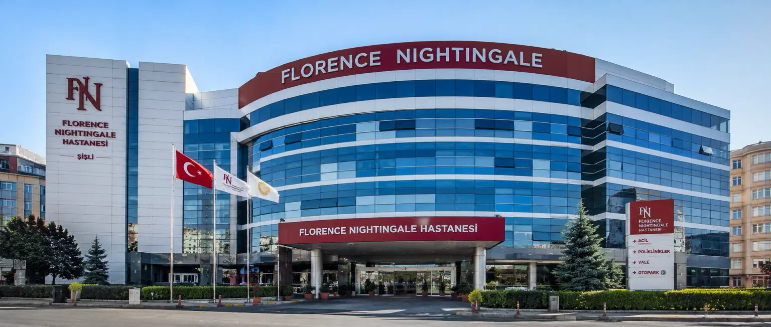
Florence Nightingale Hospital Istanbul
Abide-i Hürriyet Cd No:166, 34381 Sisli, Istanbul
Florence Nightingale Hospital Istanbul
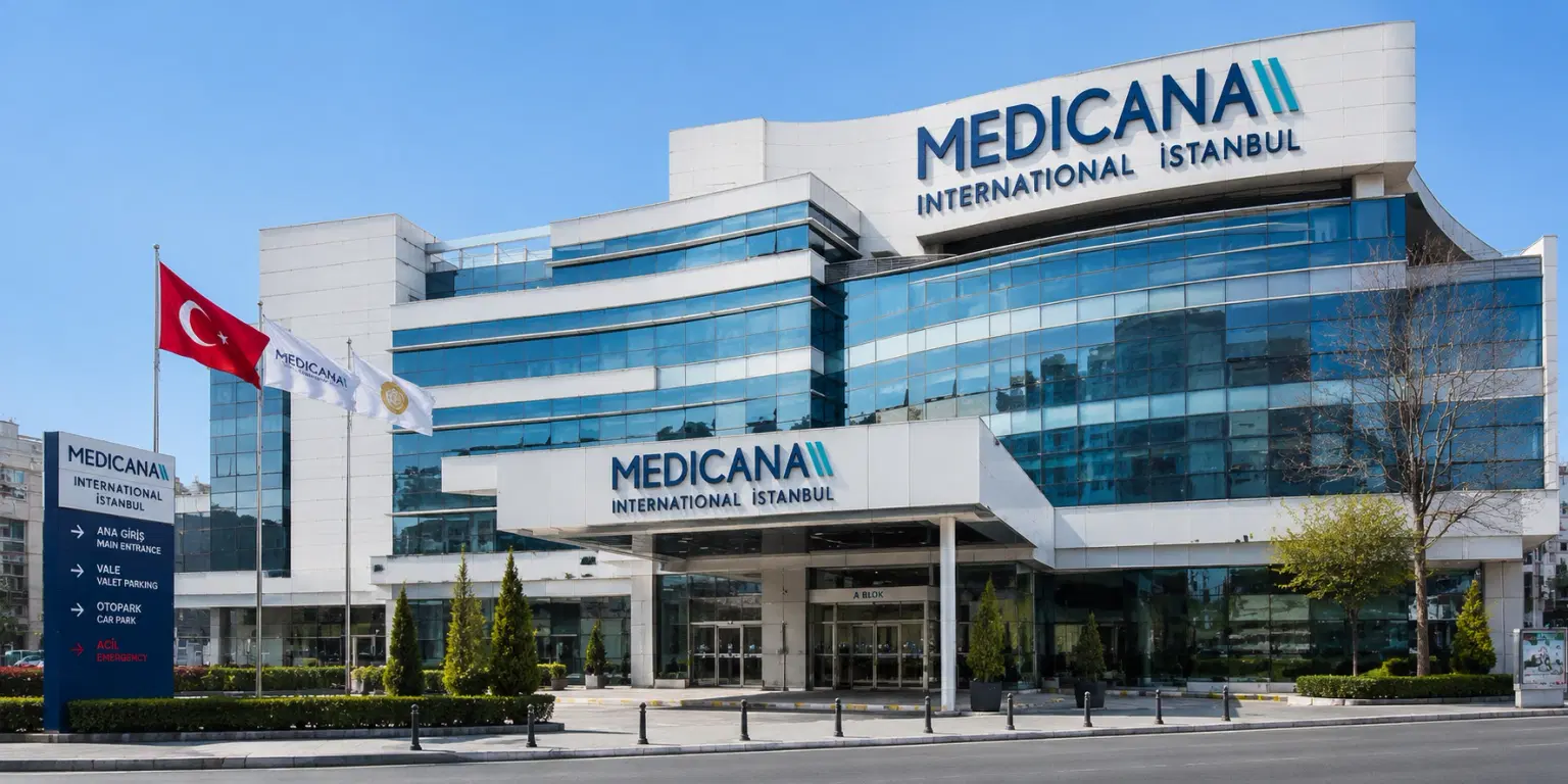
Medicana International Hospital, Istanbul
Halit Ziya Turkkani Mah. Medikal Park Cd. No:1, Beylikdüzü, İstanbul
Medicana International Hospital, Istanbul
Okan University Hospital Istanbul
Icmeler Mah. Aydınlıyolu Cd. No:2, 34947 Icmeler-Tuzla, Istanbul
Okan University Hospital Istanbul
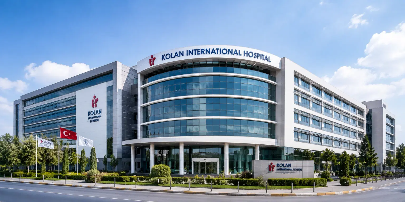
Kolan International Hospital, Istanbul
Kaptanpasa Mah. Okmeydan Kavsagi, Darulaceze Cd. No:14, 34384 Sisli, Istanbul
Kolan International Hospital, Istanbul
Meet Our Doctors
Meet our team of highly qualified and experienced medical professionals dedicated to providing the best healthcare services.

Aditi Dixit
Sr. Consultant – Women Imaging
Radiology (Specializing in Women's Imaging)
Haryana
15+ Years
Experience
Artemis Hospital
Hospital
1500
Fees

Dr. Anju Singh
Consultant - Pediatric Rheumatology
Pediatric Rheumatology
New Delhi
13+ Years
Experience
Artemis Hospital
Hospital
1500
Fees

Dr. Aparna Sinha
Associate Director
Pulmonology
New Delhi
22+ Years
Experience
Apolo Delhi
Hospital
1500
Fees

Dr. Ellora Nanda
Admin Head - Emergency
Emergency & Trauma Services
New Delhi
25+ Years
Experience
Artemis Hospital
Hospital
1500
Fees

Dr. Kamal Verma
Sr. Consultant - Radiation Oncology
Radiation Oncology
New Delhi
15+ Years
Experience
Artemis Hospital
Hospital
1500
Fees
Dr. Manish Agarwal
Director - Oncology
Oncology
New Delhi
22+ Years
Experience
Apolo Delhi
Hospital
1500
Fees

Dr Nidhi Rawal
Chief - Paediatric Cardiology
Paediatric Cardiology
New Delhi
18+ Years
Experience
Artemis Hospital
Hospital
1500
Fees

Dr. Noaline Sinha
Chairperson - Nuclear Medicine & Radio-Theranostics
Nuclear Medicine
New Delhi
15+ Years
Experience
Artemis Hospital
Hospital
1500
Fees

Dr. Priya Tiwari
Sr. Consultant
Radiology
New Delhi
12+ Years
Experience
Artemis Hospital
Hospital
1500
Fees
Dr. Rajeev Rathi
Director - Pulmonology
Pulmonology
New Delhi
34+ Years
Experience
Apolo Delhi
Hospital
1500
Fees
Booking With DIVINHEAL
Get a free consultation to understand your treatment options